Proscar
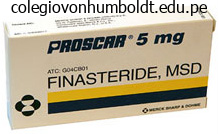
Proscar dosages: 5 mg
Proscar packs: 30 pills, 60 pills, 90 pills, 120 pills, 180 pills, 270 pills, 360 pills
Order proscar 5 mg otc
During the third trimester, ultrasound can be used to determine the placental web site precisely. However, the most important role of ultrasound in the course of the third trimester is that of figuring out fetal well-being. This is achieved by the measurement of fetal progress parameters, liquor volume and umbilical artery Doppler measurements. Prenatal diagnosis 7 Describe the strategies used for invasive prenatal analysis. Amniocentesis is the most generally used diagnostic test and could be carried out from 15 weeks to term. It is carried out in pregnancies which were recognized as excessive danger by prior screening or historical past. It is performed when a pattern of fetal blood is required, for instance to determine platelet count in alloimmune thrombocytopaenia. The purpose of any investigation of polyhydramnios is to set up a prognosis, so that a prognosis can be decided. This should embody medical historical past, as there are various diseases that may trigger fetal polyhydramnios. The most typical maternal disease associated with polyhydramnios is poorly controlled diabetes mellitus. Therefore, a random blood glucose degree ought to be obtained and this should be followed by an oral glucose tolerance take a look at, if indicated. Maternal red-cell antibodies should be checked to exclude isoimmunization, as this is associated with fetal hydrops. It is related to oligohydramnios in the different sac and requires pressing treatment by amniodrainage. Twins and higher order multiple gestations 9 Outline the issues that may happen with a twin being pregnant. Complications that occur in twins may be divided in to people who occur in all twins and people who happen specifically in monochorionic twins. Monochorionic twins have specific issues owing to the reality that the twins share the identical placenta. Uterine over-distension is assumed to be the primary reason for preterm labour in twins. In a dichorionic pregnancy, the prospect of late miscarriage is approximately 2 per cent, and for monochorionic twins the risk is as excessive as 12 per cent. The chance of poor fetal progress for monochorionic twins is almost double that for dichorionic twins. Each dichorionic twin being pregnant has at least twice the chance of a structural anomaly. Therefore, in monozygotic twins, the danger is identical as for maternal age as both fetuses arise from the identical egg. However, dizygotic twins have twice the danger, as the fetuses come from two different eggs. Monoamniotic twins have the additional risk of wire entanglement, causing fetal death late in being pregnant. Twin pregnancy may be related to assisted conception, which supplies a better danger of preterm supply. Pre-eclampsia and other issues of placentation 10 A twenty four-year-old lady presents at 36 weeks in her first being pregnant. A full history is required to ascertain whether there are any threat factors for pre-eclampsia. A detailed history of signs must be taken, notably of headache, visible disturbance and epigastric pain. An abdominal palpation will show whether the fetus is clinically small for dates. Maternal assessment ought to embrace a full blood depend to decide platelet rely. Serum uric acid levels must also be measured as these reflect both renal function and placental breakdown. A 24-hour urine assortment must be initiated to confirm that the urinary protein is > 0. An pressing mid-stream urine and microscopy must be sent to exclude a urinary tract infection.
Order generic proscar from india
Advancement of every dilator is finest completed using an alternating clockwise�counterclockwise rotational movement to minimize the chances of guidewire kinking. The shoulders of each Amplatz dilator have to be superior until completely within the entry calyx. The working sheath is introduced final, over the biggest Amplatz dilator until the leading edge of the sheath overlaps the shoulders of the Amplatz dilator (see Video 20. An further benefit of this method is offered by the relative rigidity of the dilators, which in combination with the elevated stability and tapered profile, allows higher freedom in dilation of the scarred retroperitoneum. As in the earlier system, the principle disadvantage is still the theoretical threat of extreme utility of pressure, leading to renal pelvis perforation and renal parenchymal bleeding [2, 3]. Each dilator is designed to adapt to the lumen of the subsequent successive dilator, beginning with an 8F hole guide rod. The initial hole information rod is advanced over a guidewire beneath fluoroscopy, till its tip is positioned throughout the renal pelvis. Dilation is achieved with six successive dilators, every rising in dimension by 4F, finally dilating the tract to both 24F or 26F. A metal guard at the end of every rod represents the endpoint for the insertion of the dilator [4, 5]. The main benefit of this method lies in its rigidity, which provides help for dilating through dense scar tissue. This benefit is offset by elevated potential for iatrogenic injury, due to problem with controlling the strain throughout dilation. Moreover, the need for handbook stabilization of the central rod throughout the dilation will increase the danger of perforating the renal pelvis. Modification of the standard Alken steel dilators has been described in an try to enhance the protection margin [6]. These modifications have been shown to lower trauma to the tissues and reduce the prospect of renal pelvis perforation from overadvancing the dilators. Additionally, the nonlocking mechanism allows for removal of inner dilators, permitting insertion of a ureteroscope and inspection of the tract and amassing system as the tract is being established [6]. Balloon dilator techniques the usage of balloon dilators was pioneered within the field of interventional angiography and has subsequently been tailored for use in different disciplines. The application of balloon dilation to percutaneous renal surgical procedure has been a significant development. Percutaneous nephrostomy tract balloon dilators are meant for creation of tracts in a fast, single step, eliminating the necessity for serial dilation. They are designed to be introduced in to the amassing system over a guidewire and tract dilation is carried out under fluoroscopic steering. Pressures of as a lot as 20 atm may be easily achieved with this technique, although generally, a lot decrease pressures are often sufficient for tract creation. Balloon inflation permits for full expansion of the balloon, which is adopted by insertion of a working sheath over the balloon, in a rotational method. The major advantage of the balloon dilation technology lies in the reality that the tract is created utilizing lateral, quite than angular, shearing forces. In concept, lateral forces are much less traumatic and cut back the possibility of large vessel harm and resultant hemorrhage. An further benefit is the elimination of serial dilation, which saves operating room time [7]. The dilation is also more controlled, because the balloon can simply be stabilized during Chapter 20 Dilation of the Nephrostomy Tract 235 the inflation. In earlier experience, the utilization of balloon dilators was found to lead to a decrease rate of blood transfusions compared to Amplatz and fascial dilators [8, 9]. Contemporary collection comparing the Amplatz and balloon dilation techniques have suggested the differences in issues is most likely not significant [10, 11]. The main drawback of balloon dilators are their relative greater price compared to the other systems. In a case the place more than one tract is required, the identical balloon can be utilized once more. Other considerations with the balloon catheters embrace an incapability every so often to dilate via dense scar tissue and the danger of balloon rupture inflicting barotrauma. In most conditions, the balloon dilator is superior until the proximal radio-opaque marker is positioned throughout the entry calyx (see Video 20. Correct placement of the balloon dilator is essential to provide a helpful tract and to keep away from important bleeding.
Generic proscar 5mg with mastercard
The patient in query is in her third being pregnant (G3) and has delivered three babies at >24/40 (P3). Increased cardiac output causes move murmurs within the majority of women by the tip of the primary trimester. Thereafter the rise in amniotic fluid depends on the fetal kidneys and lungs to produce fluid. Fetal swallowing helps to take away fluid and thus oesophageal and duodenal atresia are related to polyhydramnios. The periderm produces a creamy protecting layer generally known as the vernix, which rubs off after delivery. Thermoregulation, dehydration and infection, however, are all necessary issues related to the skin of untimely infants. Placental site is first evaluated at 20/40, with follow-up scans if it is low-lying. Twin pregnancy is detected on the courting scan and may be one of the best alternative to decide the chorionicity of the being pregnant. Uterine abnormalities corresponding to bicornuate uterus are often greatest seen at this stage before the cavity becomes too distended. The chorionicity of twin pregnancy is best decided in the first trimester; this should ideally occur at approximately 12 weeks. Serious fetal structural abnormalities are identified in three per cent of all pregnancies. The presence of two or more accelerations on a 20�30-minute cardiotocogram defines a reactive trace. No check ought to be performed with no full dialogue of the knowledge that the outcomes will yield and all the options that will then be obtainable. Prior to testing or prognosis, the parents should concentrate on the choices available to them. Growth restriction relates to smoking in a dose-dependent manner, therefore severe progress restriction is unlikely if smoking has been drastically reduced. Exogenous anti-D immunoglobulin is administered in an try and stop the manufacture of endogenous antibody by the mother, as this may sensitize the immune system and put subsequent fetuses in danger. The Kleihauer take a look at determines the proportion of fetal cells inside the maternal circulation and hence helps decide anti-D dosage. Studies counsel profit in ladies with a history of three or extra late miscarriages of preterm deliveries. It is finest performed after 12�14 weeks to keep away from the issues of early being pregnant loss and to assess nuchal translucency previous to intervention. A McDonald suture can usually be eliminated with out recourse to regional anaesthesia. However, if the father or mother is affected, the incidence raises to 5 per 100 stay births; therefore, all pregnant girls with congenital coronary heart disease should have a detailed fetal cardiology scan. Prophylactic antibiotics must be given to any girls with congenital heart defects. Radioactive iodine is contraindicated because of the effect on the fetal thyroid gland. The primary risks to the fetus are of development restriction, stillbirth, fetal tachycardia and untimely delivery. There is an elevated danger of hypoglycaemia, hypocalcaemia and hypomagnesaemia after start. A 24-hour urine catecholamine measurement mixed with imaging of the adrenal glands provides the analysis of phaeochromocytoma. The sudden will increase in blood pressure with headache, blurred imaginative and prescient and nervousness which are typical of 60 Obstetrics phaeochromocytoma may be mistaken for the extra common syndrome of pre-eclampsia. During normal labour steroid alternative therapy is increased to account for the extra physiological stress. Sickle cell disease is brought on by a single amino acid substitution, but thalassaemias lead to a reduced production of normal haemoglobin.
Purchase generic proscar on line
Lojanapiwat and Prasopsuk found that the supracostal and subcostal approaches had been both efficient with acceptable complication charges [14]. Other proposed advantages include avoiding the increased value and studying curve associated with the versatile instrumentation required within the single tract strategy [19]. Large stone burdens may additionally be approached with a single entry tract as a outcome of the technologic advances in flexible instrumentation, lasers, and basketing units. This approach has been described by several endourologists [20] and is presently the procedure of selection at our institution. Usually this is through the higher pole calyx, which allows the surgeon to entry the overwhelming majority of the collecting system. The commonest entry websites are both below the 12th rib or between ribs 11 and 12. After obtaining entry and tract dilation, inflexible nephroscopy is utilized to clear as much stone burden as attainable, while minimizing renal parenchymal trauma. The commercially available devices embrace ultrasonic and pneumatic units, or a mix of the 2. The benefit of the mixture lithotripters is their capacity to fragment stones of various compositions while concomitantly evacuating stone particles. Calculi which are small enough are eliminated with a stone basket through a flexible cystoscope or ureteroscope. Larger or impacted stones are fragmented with the holmium laser and then both eliminated with a stone basket or positioned so they can be treated with the inflexible nephroscope. At the conclusion of the process, we routinely depart either a single or double J ureteral stent. Historically, a large bore nephrostomy tube (22F council-tip catheter) was routinely placed to externally drain the kidney, maintain percutaneous access, and tamponade renal parenchymal bleeding. It is important to think about that complete stone removing could not all the time be achieved regardless of perceived on-table stone clearance, and sustaining the tract with a nephrostomy tube for re-assessment nephroscopy may be helpful. In a sequence of 45 renal items with a stone burden of 5 cm or greater undergoing single tract access, Wong and Leveillee reported a stone-free fee of 95%. If residual stone is present, and the affected person was left with a nephrostomy tube to maintain entry, second look nephroscopy is carried out on postoperative day 2 throughout a single hospital admission. If the affected person was left tubeless, and residual stone is discovered, it may be approached with second-look retrograde ureteroscopy during the same hospitalization or at a later date depending on affected person or surgeon choice. Percutaneous access utilizing ureteroscopic help Large stone burdens could make acquiring access tough as there may not be enough house to enter and safely dilate a tract. If entry could be achieved, and a tract is safely dilated, it may nonetheless be tough to reach the entire intrarenal collecting system via a single tract despite the usage of versatile instrumentation. To tackle these issues, retrograde ureteroscopy has been utilized to establish the ideal calyx for percutaneous entry [28, 29]. Ureteroscopy could be carried out initially to assess the stone burden/location and to endoscopically clear a calyx to present optimum access. Once recognized and any obstructing stone burden cleared with retrograde lithotripsy, the access needle could be positioned and confirmed under direct ureteroscopic vision [30]. Use of a ureteral access sheath can facilitate retrograde lithotripsy, which may be performed with the affected person in either the supine or susceptible position. Supine versus susceptible percutaneous nephrolithotomy Recently, there was interest within the optimal affected person positioning to maximize stone-free charges whereas minimizing the danger for position-related issues. Proponents of the supine position stress the potential anesthesia benefits, particularly in obese sufferers, the place the prone place can limit respiratory capabilities. Other perceived advantages include improved patient comfort and elevated versatility of stone manipulation. Access is often subcostal via a posterior decrease pole calyx, which minimizes the danger for pleural harm. In addition, it has been stressed that by maintaining the supine place, retrograde ureteroscopy can be carried out in addition to normal antegrade therapy. Their results led the authors to conclude that this approach was both secure and efficient with an overall complication rate of 38. Proponents of the susceptible position give attention to the familiarity of the affected person anatomy as properly as decreased renal mobility facilitating percutaneous access. Using a splitleg desk, the affected person may be positioned to simply carry out flexible cystoscopy and access the ureter. Prone endoscopy has a steep, but easily overcome, learning curve, and this talent can simply be obtained by any endourologist familiar with flexible instrumentation.
Buy discount proscar online
The supracostal percutaneous nephrostomy for remedy of staghorn and complex kidney stones. Complications associated with percutaneous nephrolithotripsy: Supra- versus subcostal entry. Is the 10th and 11th intercostals area a safe strategy for percutaneous nephrostomy and nephrolithotomy Impact of percutaneous entry point number and site on complication and success rates in percutaneous nephrolithotomy. Upper-pole access for percutaneous nephrolithotomy: Comparison of supracostal and infracostal approaches. Risks, benefits, and problems of intercostals versus subcostal method for percutaneous nephrolithotomy. This low incidence is likely the end result of the colon hardly ever being retrorenal (reported in roughly zero. When ninety sufferers have been studied in the inclined place, a retrorenal colon was present in 10%. The place of the colon is often anterior or anterolateral to the lateral renal border. Therefore, threat of colon harm normally exists solely with a very lateral (lateral to the posterior axillary line) puncture. Ultrasound-guided renal percutaneous entry could be carried out in sufferers at elevated danger of having a retrorenal colon [19]. Diagnosis Prompt, early recognition of a colonic perforation is crucial to restrict critical infectious penalties. Colonic perforation must be suspected if the patient has intraoperative or instant postoperative diarrhea or hematochezia, indicators of peritonitis, or passage of gas or feces by way of the nephrostomy tract [21]. Otherwise, postoperative nephrostography earlier than nephrostomy removing can reveal the presence of colonic contrast. Antibiotic therapy, fluid resuscitation, and even the administration of pressors may be essential to treat these patients. Risk components Displacement of the colon posterior to the kidney increases the chance of colon perforation, and is seen in aged sufferers with chronic constipation or sufferers with other causes of colonic distention, patients with previous main belly surgery (jejunoileal bypass, partial jejunoileal bypass), neurologic impairment, and institutional bowel resulting in an enlarged colon. In case of intraperitoneal colonic perforation, peritonitis, sepsis, or failed conservative administration, open surgical exploration ought to be performed, and a colostomy is normally essential. Some authors have reported on the nonoperative administration of a nephrocolic fistula that resulted from percutaneous nephrostolithotomy with adequate urinary and colon drainage, elemental food regimen, and antibiotic protection. The affected person should be given broad-spectrum antibiotics or triple antibiotic coverage and be on a low-residue food regimen. This permits the renal amassing system to heal and the medial colonic wall to close. After 5�7 days, if the colostogram or a retrograde pyelogram reveals neither extravasation nor colonic communication with the accumulating system, the Foley catheter is removed and the colostomy tube withdrawn however nonetheless stored as a drain exterior the colon. Colonic damage during supine percutaneous nephrolithotomy (courtesy of Pr Bertrand Dore). Chapter 32 Bowel and Other Organ Injury during Percutaneous Renal Surgery 351 the scientific signs had been rectal hemorrhage with shock in a single case and passage of gasoline through the nephrostomy tract in the different. Three sufferers have been thought-about lean, and the other two were of common physique habitus. Recognition of colon injury occurred postoperatively in four sufferers and intraoperatively in a single patient. Presenting signs and signs included fever, fecaluria, belly ache, and leukocytosis. The authors concluded that retroperitoneal colon accidents can be efficiently managed conservatively with early recognition and acceptable drainage of the urinary and intestinal tracts. They additionally really helpful, for high-risk patients, a more superior and medial puncture. The authors evaluated operative particulars and postoperative course to decide the time and mode of prognosis of colonic damage, and remedy strategies and outcome.
Syndromes
- Sunken appearance to the eyes
- Damage to nearby structures
- Avoid chewing gum or sucking on candies
- Headache
- You should be able to return to your regular activities the next day.
- Throat culture
- Smoking
- Fever higher than 101°F (38.3°C)
- Infertility (if both testicles are removed)
- Infection (a slight risk any time the skin is broken)
Buy generic proscar on line
Swabs must be taken for chlamydia, gonorrhoea and different sexually transmitted ailments, and a smear must be obtained if affected person has not had one as a part of the traditional recall process. One should observe age, historical past of any kids on this relationship or other relationships, smoking, alcohol use and occupation. It is necessary to enquire about testicular trauma, undescended testes, mumps and sexually transmitted ailments. On examination, one ought to assess the dimensions of each testis, examine for varicoceles and descent, and notice secondary sexual traits. Disorders of early pregnancy 7 Write quick notes on threatened miscarriage, silent miscarriage and incomplete miscarriage. The patient presents with vaginal bleeding that could be associated with suprapubic pain. Ultrasound demonstrates a gestational sac with a fetal pole and the fetal coronary heart is seen. Sometimes the analysis is made incidentally at ultrasound scan when sufferers come for a routine 12-week or 20-week scan. The analysis is confirmed if ultrasound reveals an embryo of <20 weeks with no fetal heart and no indicators of expulsion. The management could be either expectant, surgical evacuation or medical, and is decided by the size of the products of conception throughout the uterine cavity. Clinical presentation the majority of sufferers current with belly ache � vaginal bleeding. Occasionally, sufferers have shouldertip pain indicative of free blood in abdominal cavity. Benign illnesses of the uterus and cervix 9 Write short notes on the rules of a screening programme. There are 10 ideas of screening that are actually adopted by the World Health Organization. Endometriosis and adenomyosis 10 Outline the 4 theories for the pathophysiology of endometriosis. One concept is that endometriosis occurs on account of retrograde menstruation and that implantation of endometrial glands and tissue happens in to the peritoneal surface. Another concept is that peritoneal cells and cells in the ovary that are derived from the M�llerian duct bear dedifferentiation again to their primitive origin after which transform in to endometrial cells. Some ladies of sure genetic/immunological predisposition might possess elements that render them prone to the development of endometriosis. Occasionally endometriosis may be discovered inside and outside the peritoneal cavity, such as skin, kidney and lung. This may happen because of embolization of endometrial tissue by way of vascular lymphatic channels or at surgery. History the salient features embrace dysmenorrhoea, the demonstration of cyclical pelvic pain, deep dyspareunia, a historical past of sub-fertility or infertility. Bladder symptoms might embrace cyclical haematuria or ureteric obstruction, and bowel symptoms could embrace cyclical rectal bleeding or pain on defecation. It is typically attainable to palpate nodules of endometriosis on rectovaginal examination. A diagnostic laparoscopy with or with out tubal patency testing will confirm a prognosis of endometriosis, and endometrial explants may be seen throughout the peritoneal cavity. Adenomyosis tends to have an effect on ladies between the ages of thirty and forty, whereas endometriosis tends to have an result on women in their late twenties and thirties. In girls with an endometrioma, tenderness is often elicited in the pouch of Douglas, rectovaginal septum and adnexae. Magnetic resonance imaging supplies further enhanced photographs and is the investigation of selection. If the affected person has endometriosis, ultrasound could demonstrate the presence of endometriomas.
Cheap proscar on line
Other organ injury Though less widespread than pulmonary problems, injuries to adjoining organs, together with the liver, spleen, and intestines, can not often occur throughout upper calyceal access. A retrorenal left colon, occurring in 10% of patients within the susceptible place, could prohibit entry to the tenth or 11th intercostal area with increased danger associated with medial punctures [26]. Additionally, a supra11th entry might puncture the liver in 14% and spleen in 33% of patients, notably throughout inspiration [7]. In hemodynamically stable patients, liver accidents can typically be managed conservatively with tube drainage and serial monitoring [27]. Splenic accidents, nevertheless, are associated with elevated bleeding and should require instant exploration and splenectomy [29]. During needle placement, medial puncture by way of the paraspinal muscular tissues is related to elevated ache and should be averted if possible [3]. Several suggestions have been made to decrease pain following upper pole entry. Supra-12th rib puncture should be approached lateral to the erector spinae muscular tissues towards the superior portion of the rib throughout maximal expiration. Intraoperative fluoroscopy of the lung fields should be performed at the conclusion of the procedure, and a powerful suspicion for pulmonary accidents should be maintained within the postoperative interval. Patients ought to be endorsed appropriately relating to the potential risks and complications when higher calyceal access is anticipated, notably almost about intrathoracic problems. However, with consideration given to anatomic concerns, supracostal access to the higher pole could be obtained safely and efficiently to contribute to profitable percutaneous renal surgical procedure. Approaches to the superior calyx: Renal displacement approach and evaluation of choices. Prospective evaluation of safety and efficacy of the supracostal approach for percutaneous nephrolithotomy. Safety and efficacy of supracostal access in tubeless percutaneous nephrolithotomy. Risks, advantages and complications of intercostal vs subcostal method for percutaneous nephrolithotripsy. Splenic damage: rare complication of percutaneous nephrolithotomy: report of two instances with evaluate of literature. Using and selecting a nephrostomy tube after percutaneous nephrolithotomy for large or complicated stone illness: a remedy strategy. Use of lower pole nephrostomy drainage following endorenal surgery via an upper pole access. Nephrostomy tube after percutaneous nephrolithotomy: Large-bore or pigtail catheter Tubeless and stentless percutaneous nephrolithotomy in sufferers requiring supracostal entry. The rapid dissemination of image-guided methods in urology and interventional radiology has contributed to the innovation of percutaneous renal procedures. Simple drainage has advanced in to percutaneous therapies for stone disease, higher tract and renal cancers, and urinary diversion/drainage, amongst others. The advancement of endourologic strategies has led urology in to the minimally invasive era. This speedy innovation has pressured urologists to become proficient in percutaneous renal procedures, offering optimal therapy for the increasing number of pathologies amenable to percutaneous treatment. On uncommon events, access to numerous imaging modalities could also be unavailable, or proficiency with image-guided percutaneous nephrostomy placement could additionally be lacking. Anatomy (see also Chapter 6) the retroperitoneal location of the kidney allows for relatively protected percutaneous access by way of a posterior method. Nephrostomy placement without image steering is accomplished using anatomic landmarks, assuming regular renal anatomy. A thorough understanding of renal anatomy and relations is crucial to keep away from issues and gain access to the accumulating system [8]. The kidney is a mobile organ and its place can range depending on patient position and phase of respiration. With respiratory excursion, the kidneys descend on average 4�5 cm, reaching as far as the iliac crest. Generally, the best kidney lies 1�2 cm decrease than the left because of its place beneath the liver.
Generic 5mg proscar
This scope was primary in design, consisting of a direct-vision hollow tube via which candlelight was transmitted by a mirror. Gustave Trouv� additional upgraded indirect illumination by candlelight to electrical illumination utilizing glowing heated platinum wires at the tip of the instrument in 1873 [5]. Year 1806 1853 1873 1879 Sixties Nineteen Sixties Pioneer Phillip Bozzini Antoine Jean Desormeaux Gustave Trouv� Maximilian Nitze and Joseph Leiter Several contributors Harold Hopkins Rigid endoscope and technologic advances German physician who launched the ""Lichtleiter" or gentle guiding instrument French surgeon who first introduced the "endoscope". The NitzeLeiter cystoscope, a rudimentary version of the modern cystoscope, was constructed in 1879, incorporating a easy lens system in to the viewing tube [5]. The introduction of fiberoptic lighting in the Nineteen Sixties replaced the incandescent bulb, revolutionizing the manufacture of rigid endoscopes. The design was primarily based on the principle of whole inside reflection; when light travels in a transparent medium such as glass, inner reflection of the light occurs at the interface between the medium and its surroundings, as demonstrated by John Tyndall in 1854 [6]. This bodily property of internal reflection permits the "bending" of light inside versatile glass. Thus, gentle travelling inside a small-diameter flexible glass fiber surrounded by cladding of lower refractory index could be transmitted over an extended distance with minimal degradation. The benefit of modern fiberoptic lighting is larger illumination by cooler light, ultimately making it safer. An additional benefit is that the scopes could be made with a smaller-profile shaft, allowing more room throughout the shaft for the addition of an irrigation and instrument channel. The fiberoptic cable is usually connected through a lightweight publish to the endoscope, however might alternatively be inbuilt to the design of the scope. The revolutionary work of the British physicist Harold Hopkins led to the subsequent main breakthrough in rigid endoscopic design with the introduction of the rod�lens system in the Nineteen Sixties [7]. Until then, the shaft of the scope consisted of a hollow tube with a series of thin relay lenses separated by long air spaces. The relay lenses needed to remain in precise alignment and any displacement of the lens resulted in a big loss of picture transmission. Hopkins replaced the thin relay lenses within the shaft of the endoscope with lengthy, contoured glass rods acting because the transmission medium, whilst the thin pockets interspersed between the glass rods acted as lenses. With the telescope primarily being Chapter 34 Rigid and Flexible Ureteroscopes: Technical Features 367 made of glass, which has a higher refractive index than air, light transmission could presumably be increased ninefold over earlier lens techniques, with the additional advantage of decreased image distortion and a wider viewing angle. The outer diameter of the endoscope shaft is also lowered, paving the way in which for the development of the rigid ureteroscope and the introduction of working channels. The old lens system was notorious for its lack of sturdiness and the Hopkins rod�lens system produced a big improvement on this space. There had been a few apparent weaknesses in the rod�lens design: any deviation in the use of the cystoscope from being used in straight strains, as can occur with the application of torque to the shaft, may lead to misalignment of the rod�lens arrange and the permanent disappearance of up to half the image. The downside manifests itself as a halfmoon or crescent-shaped defect when viewing by way of the scope eyepiece. This design flaw would be seen to be disadvantageous in the passage of the endoscope by way of the pure undulations of the ureter. A further consideration for ureteroscopic design facilities on the diameter of the lens that dictates the dimensions, or diploma of magnification, of the picture; the reduced diameter of the ureteroscope would inadvertently have a smaller picture than the bigger cystoscope. Endoscopic evolution Considering that Young undertook the first recorded ureteroscopy in 1912, it was one other sixty five years earlier than Goodman [8] and Lyon et al. Goodman reported utilizing an 11F pediatric rod�lens cystoscope to inspect the distal ureter in three adults [8]. In one of these patients, a distal ureteral tumor was fulgurated, marking the first ureteroscopically handled tumor. They were able to dilate the orifice to 16F, allowing insertion of a normal length 13F cystoscope for distal ureteroscopy in males. These early investigators demonstrated the benefit and safety of inflexible ureteroscopy for the distal ureter in both sexes. The first rigid ureteroscope particularly designed for ureteric use was produced in 1979 by Richard Wolf Medical Instruments and was modeled on a pediatric cystoscope. The 13F sheath was used only for inspection, however the bigger sheaths allowed simultaneous passage of a ureteral catheter or basket for stone manipulation and removing. For the primary time, ureteral calculi have been visualized, engaged in a basket, and eliminated [10, 11].
Generic proscar 5 mg online
Several techniques can be found that can be applied to the treatment of upper tract neoplasms ureteroscopically. These techniques, which might take away or ablate tissue within the upper tract using inflexible or versatile ureteroscopes, embody mechanical removal, electrosurgical resection, fulguration, and laser remedy. These are thought-about in greater detail because of their significance in treating neoplasms inside the higher urinary tract. The strategies used for the ureteroscopic biopsy of tumors result in the removal of tissue quantity. A flat wire basket can take away several millimeters of papillary tumor with each software (see Video forty one. Although the cup biopsy forceps take away a small volume with each bite, repetitive sampling can remove a small tumor (see Video forty one. The base of the tumor can then be coagulated with one of many different devices (see Video forty one. Electrosurgical methods similar to these used in the bladder have been utilized for small distal ureteral neoplasms. Historically, resection was the primary to be used for therapy of upper tract neoplasms [58, 59]. Rigid ureteral resectoscopes, much like a protracted model of a pediatric resectoscope, can be utilized in a similar way to different resection procedures for tumors in the distal ureter. The loop takes a small chunk of tissue and it may become necessary to clear the loop earlier than eradicating the following piece of tissue. Extreme care should be taken to keep away from resecting via the full thickness of the ureteral or renal pelvic wall, and likewise to avoid fulgurating a big area of the ureter, which can lead to scarring and stricture formation [60]. Chapter 41 Ureteroscopic Diagnosis and Treatment of Upper Urinary Tract Neoplasms 443 laser can penetrate to a depth of roughly 5�6 mm, it could not have an effect on the entire depth of the tumor. Coagulated tissue is removed with a grasper or basket to expose portions of the tumor in any other case not seen ureteroscopically. It is a solid-state pulsed laser that may fragment calculi, and coagulate, ablate, and take away tissue. This laser produces mild at a wavelength of 2100 nm, which could be carried alongside low water content material fiber. This laser is particularly useful for ureteral lesions since it can ablate and remove a visually occlusive neoplasm to open the lumen for inspection more proximally [65�67] (see Video 41. The laser fiber have to be placed in touch with or very near the tissue to be handled. It is commonly essential to discontinue treatment to enable the sphere to clear, since appreciable debris is fashioned throughout therapy. Bleeding occurs occasionally and could be controlled better at decrease energies or by moving the fiber barely away from the tissue to diffuse the laser beam and enhance coagulation. There can be scientific evidence that an extended pulse length, such as seven hundred ms, quite than the 350 ms usually used for lithotripsy, will also improve coagulation (see Video forty one. The very restricted penetration permits precise management and the laser, thus, can be used for lesions positioned on the level of the iliac vessels and the renal pelvis close to the renal vessels. Great care is employed to avoid ablation and resection by way of the wall of the ureter or renal pelvis itself. The argon laser has major theoretical advantages for endoscopic treatment as nicely. Wavelengths of 488� 514 nm have been used to deal with superficial bladder most cancers and likewise ureteral neoplasms. The penetration is restricted to 1 mm and the laser gentle may be delivered with a fiber of 300 or 600 m diameter. Johnson reported treating tumors with continuous wave argon laser energy with a quartz fiber, utilizing a contact mode with the ability set at 5 W [68]. There was passable ablation of tumors in all three sufferers, but other confirmatory reviews are nonetheless not available. The techniques used for therapy, the situation and traits of the tumors, and follow-up strategies and duration have diversified widely. There were asynchronous recurrences in three patients which remained low grade and no patient required surgical remedy in the course of the research interval.
Cheap proscar online visa
The point of obstruction was identified in all patients, and different etiologies for obstruction. The disadvantages of this method, in addition to the lack to identify the ureteral calculus, are the image acquisition time of 34 min and the worth of the procedure. Again, for patients with allergic reactions to iodinated distinction and renal failure, this technique may have its place. The experimental models for ureteral obstruction embrace: (1) unilateral or bilateral ureteral ligation; (2) burying the mid ureter within the psoas muscle; and (3) placing silastic plastic tubing around the ureter to constrict the ureteral lumen. Hemodynamically, part 1 is characterised by an preliminary afferent arteriole vasodilation [6] followed by an efferent arteriole vasoconstriction in phase 2 and afferent arteriole vasoconstriction in phase three [32]. Utilizing the injection of microspheres during ureteral occlusion, they Chapter 7 Pathophysiology of Urinary Tract Obstruction Table 7. Kessler demonstrated a concentrating defect in the obstructed kidney within 5�8 min of ureteral occlusion [45]. The three phases are designated by Roman numerals and divided by vertical dashed traces. Wilson concluded that volume expansion was enhancing an already-present defect in water reabsorption either in distal nephrons and/or deep nephrons or in amassing ducts of the obstructed kidney. Hanley and Davidson additionally described an incapability of the kidney to concentrate urine after obstruction [48]. Chapter 7 Pathophysiology of Urinary Tract Obstruction the characterization of the aquaporins, a household of membrane water channels, offers a molecular foundation for transmembrane water motion [50]. Aquaporin-2 is the predominant vasopressin-sensitive water channel of the accumulating duct. Consistent with the decreases in aquaporin was a significantly increased free water clearance in the obstructed kidney. The postobstruction kidney has the next bicarbonate reabsorption price than the contralateral kidney [52]. This is accompanied by a twofold larger fractional excretion of sodium within the obstructed kidney than in the control kidney. However, the decrease in phosphate excretion is believed to be secondary to a decreased filtered load of phosphate. The increased excretion of salt and water is secondary to a decreased reabsorption within the distal nephron without a concomitant lower in proximal reabsorption [54]. This change in intratubular stress brought on a decrease within the hydrostatic pressure gradient from 31. There was little change in glomerular capillary pressure (46 and 50 mmHg, pre- and postobstruction, respectively). Wilson and Honrath demonstrated such a substance with crosscirculation research in rats [62]. The urinary flow extra closely adopted the osmolar excretion than the sodium excretion in this patient study. The predominant urine solute during this osmotic diuresis was urea, accounting for 37�68% of the whole urine osmolality. They found that patients with chronic obstructive uropathy had an increase in whole exchangeable body sodium before launch of obstruction. After aid of the obstructive uropathy, whole exchangeable body sodium ranges returned to regular inside 2�3 weeks. There was a rise in each absolute and fractional excretion of sodium that subsided with enchancment in renal function. Standard error of mean value is proven; significance of the distinction from the mean control value: (reproduced from Wilson and Honrath [62], with permission). They observed impaired fractional sodium reabsorption in the distal tubule, with normal fractional reabsorption of sodium within the proximal tubule. Jaenike did show a defect in sodium transport in the distal tubule and instructed that this defect is a direct mechanical impact of obstruction at the distal tubule.
Real Experiences: Customer Reviews on Proscar
Kaffu, 24 years: The use of ureteral stents and new and future applied sciences may even be discussed. It is of notice, and not surprising, that the retreatment charges were highest with the piezoelectric system, ranging between 14% and 51% and averaging 27%. Increasingly, issues associated to using anticoagulant drugs prior to urologic surgical procedures have arisen. The technique is predicated on animal studies by Davis in 1943 with intubated ureterotomy [49].
Khabir, 51 years: The ultrasound-guided approach is protected with a really low danger of major problems. Why massive stones, albeit satisfactorily disintegrated, are associated with significantly extra residual fragments is less simple to perceive. If signs are minimal and the pneumothorax is small, no other therapy is critical. Guidewires Guidewires are key to a profitable endoscopic procedure because they allow constant access in to the upper tract.
Daro, 50 years: For example, the problem of the open water tub was addressed by enclosing the shock head and through the use of a rubber membrane to couple the shock wave to the body. This mixture of inflexible and versatile ureteroscopy supplies complete visual inspection of the entire upper urinary tract. Thalamus � Sensory "relay station" between lower-order afferents and the cortex Clinical note: Thalamic (pain) syndrome is a uncommon condition in which destruction or ischemia of the thalamus results in hypersensitivity to a wide selection of stimuli. Rigid (rod�lens) cystoscopes supply higher image quality and irrigation capabilities, and are simpler to operate than commonplace versatile tools.
8 of 10 - Review by J. Osmund
Votes: 321 votes
Total customer reviews: 321
References
- Gelinas C, Johnston C. Pain assessment in the critically ill ventilated adult: validation of the Critical-Care Pain Observation Tool and physiologic indicators. Clin J Pain. 2007;23(6):497-505.
- Van Sickels JE, Tiner BD, Alder ME. Condylar torque as a possible cause of hypomobility after sagittal split osteotomy. Report of three cases. J Oral Maxillofac Surg 1997;55:398-402.
- Damiano, R., Autorino, R., De Sio, M., Giacobbe, A., Palumbo, I.M., D'Armiento, M. Effect of tamsulosin in preventing ureteral stent-related morbidity: a prospective study. J Endourol 2008;22:651-656.
- Taft C, Karlsson J, Sullivan M. Do SF-36 summary component scores accurately summarize subscale scores? Qual Life Res 2001;10(5):395-404.


