Fucidin
Fucidin dosages: 10 gm
Fucidin packs: 1 creams, 2 creams, 3 creams, 4 creams, 5 creams, 6 creams, 7 creams, 8 creams, 9 creams, 10 creams
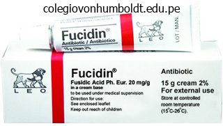
Generic 10 gm fucidin amex
Most bronchoscopists prefer to maintain the patient spontaneously breathing in case the overseas body turns into dislodged during the endoscopy. The use of the pulse oximeter is critical when assessing air flow and oxygenation. Positive mask induction with ventilatory strain should be applied smoothly and, ideally without inciting respiratory tree irritability that leads to bucking or coughing, which can dislodge the international body. After secure induction of common anesthesia, with the patient spontaneously breathing, the bronchoscopist is supplied entry to the airway. Prompt direct laryngoscopy is performed by the bronchoscopist, followed by insertion of a rigid ventilatory bronchoscope. The inflexible ventilatory bronchoscope allows for simultaneous inhalation of oxygen and anesthetic gases, and permits immediate removal of thickened secretions by suctioning. Diagnostic examination of the larynx and higher trachea is achieved by concurrent insertion of a rod lens optical telescope for magnification and transmission of the anatomy to the video screens in the working room suite. The operative examination entails careful inspection of the entire tracheobronchial airway. After advancing the bronchoscope slowly to the carina of the trachea, the bronchoscopist should focus on removing of all secretions, and careful inspection of all secondary bronchi. Bleeding from granulation tissues can turn out to be a hard problem and is finest managed through the instillation of vasoconstrictor options, corresponding to oxymetazoline or epinephrine (1:10,000 or stronger), to guarantee adequate publicity of a chronically lodged foreign physique earlier than any try is made for removal. In some situations, the main edge may be rotated if deemed necessary to grasp for extraction. Peanuts cause important airway irritation and edema and can fragment during manipulation and suctioning. Steroids are frequently given to reduce edema from endoscopic instrumentation trauma. Most sufferers can be discharged inside 24 hours of remark after surgery if the interval of remark suggests no irregular respiratory symptoms. Follow up bronchoscopy may be essential to assess for underlying bronchi or trachea for signs of stenosis or subsequent stricture improvement. Delicate debridement of residual granulation tissue with forceps or balloon dilation of the affected bronchus could also be necessary to forestall stricture. The elevated frequency of esophageal foreign bodies is attributable to each the truth that objects placed inside the oral cavity will set off the swallow mechanism, and the unsafe "eating habits" of very younger children. Presentation Most esophageal overseas our bodies cause minor signs of dysphagia, odynophagia, drooling, vomiting, and pain. Fever, hemoptysis, hematochezia, or neck crepitus are extra worrisome presenting manifestations. In rare circumstances, longstanding bronchial foreign our bodies can erode through the tracheal wall into the esophagus. In cases of tracheosophageal perforation, subcutaneous emphysema can be an additional manifestation. Button batteries carry a high electrical cost that may harm the mucosa and promptly lead to perforation of the esophagus. The increasing appearance of these small batteries in digital gadgets similar to remote controls, watches, greeting cards, and calculators has resulted in a dramatic enhance of their aspiration. Button batteries lodged within the esophagus must be treated aggressively with instant endoscopy and removal because of the high rate of mucosal injury and subsequent perforation if they remain lodged in the esophagus for even just some hours. The batteries conduct electrical currents, which shortly destroy adjacent tissues. Current battery models are more powerful than previous era batteries and thus extra damaging. Chest movies should be rigorously scrutinized to assess for changes within the periphery of the disc battery, which differentiates batteries from coins, since batteries have a "double ring" look. During inflexible esophagoscopy, the esophagoscope should be advanced using direct imaginative and prescient of the esophageal lumen. Injury to the esophageal mucosa should be famous and documented as strictures can develop.
Syndromes
- Calluses and corns: Thickened skin from rubbing or pressure. Calluses are on the balls of the feet or heels. Corns appear on the top of your toes.
- How often do you bathe or shower?
- Endocardial cushion defect
- Swallowing difficulty
- Voice change (deepening)
- Different joints in the body may become hard to move. The shoulder and other joints may dislocate.
- Cervical warts (infection with human papilloma virus, or HPV)
- Lowering salt in your diet (no more than 1,500 mg/day of sodium)
- Difficulty breathing (rare)
- Patent ductus arteriosus (PDA)
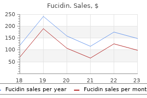
Buy generic fucidin 10gm on line
The septum is a universal landmark that presents as a whitish, comparatively avascular surgical aircraft easily recognized from the overlying orbicularis muscle. While the septum rarifies with age to produce a herniated look in some patients, in other patients getting older modifications cause atrophy and inferior migration of the superior orbital fats to produce superior periorbital hollows. The higher eyelid fats pads and the lacrimal gland lie posterior to the orbital septum. The trochlea separates the medial (nasal) pad from the central (preaponeurotic) fat pad and trochear trauma throughout blepharoplassty may lead to superior indirect dysfunction and a Brown syndrome. The medial and central fats pads differ in appearance with the former showing pale white and the latter displaying a extra yellow colour. The medial palpebral artery usually lies superficially and can be cauterized prior to transection. The lacrimal gland extra laterally, nevertheless, accommodates branches of the lacrimal artery, which produce extra bleeding when transected. Preaponeurotic fat excision often merely hastens the hollowing of the superior sulcus, which may impart a more aged appearance quite than a rejuvenated one. Some patients profit from volume augmentation of the superior periorbital hollows, quite than fats debulking. The medial-most central fats should always be spared to forestall a localized sunken area over the trochlea. Upper Blepharoplasty Procedure Topical anesthetic drops are placed in both eyes as a end result of the povidone iodine solution utilized later irritates the ocular floor. The upper lid is infiltrated with a mixture of 1% lidocaine with 1:100,000 epinephrine, hyaluronidase, and bicarbonate resolution using a 30-gauge needle. The patient is prepped and draped within the usual sterile trend for facial surgical procedure and eye protectors are placed. The marking for the blepharoplasty incision begins by placing the eyelid on stretch manually to expose the pre-tarsal eyelid tissues. When levator aponeurosis dehiscence considerably elevates the peak of the eyelid crease, a central height of roughly eight mm in males and approximately nine mm in ladies may be used somewhat than marking the elevated eyelid crease. When the crease is uneven, the decrease of the two values must be used on each side to create incision traces at the identical top on each eyelid. The lateral marking extends past the lateral canthus and turns into extra horizontal or angles barely upward alongside the relaxed skin rigidity lines. The medial marking extends no additional than the higher punctum and angles superiorly for the final two to three mm when medial cutaneous redundancy exists. After the inferior incision design is then repeated on the contralateral side the markings must be in contrast for symmetry. Calipers assist to assess symmetry by measuring the gap from the brow to the superior marking and by measuring the amount of pores and skin within every marking. The superior marking should typically arc wider laterally and taper narrower medially to account for the surplus redundant tissues in the lateral portion of the eyelid. After skin flap excision, light cautery provides hemostasis from bleeding vessels, which generally lie inside the orbicularis oculi. If blepharoplasty requires orbital fat removing, the orbital septum is opened by elevating the septum with forceps and slicing the septum perpendicularly with Westcott scissors, laser or electorcautery. This fills the preaponeurotic house and brings the septum away from the levator, buffering levator from injury upon opening the septum. The septum can also be elevated with forceps during incision to keep away from levator injury. The central flap ought to contain only skin or skin and orbicularis muscle as determined by the preoperative plan. Some surgeons use interrupted sutures, especially medially to allow for some granulation in this space to reduce medial web formation. The affected person is instructed to restrict actions, use only acetaminophen with or with out opioid analgesics, and apply ophthalmic antibiotic ointment to the injuries four occasions daily.
Discount 10 gm fucidin amex
The eburnated kind, also identified as the compact kind, consists of dense bone, and lacks Haversian canals. Common signs and indicators include frontal ache and headache, as nicely as sinusitis and/or mucocele formation from obstruction of adjoining sinus ostia. These lesions are often discovered to be pedicled to comparatively normal showing adjacent bone although in depth lesions might cause some bone-thinning secondary to the mass impact. This permits choice making and surgical intervention previous to the development of problems, should a lesion enhance in dimension on serial imaging. Rapid growth, infection, compression of vital constructions, extreme ache, facial deformity, vision changes, mucocele formation, and intraorbital and/or intracranial complications are all indications for tumor resection. These include a tumor occupying higher than 50% of the frontal sinus,eighty three a posteriorly based mostly frontal sinus lesion,87 and any osteoma occupying the frontal recess or ethmoid sinus cavity. In basic, approaches can be divided into three teams: endoscopic, open/external, or combined. Regardless of the strategy chosen, the targets of surgery should include complete tumor removing with minimal damage to surrounding mucosa and vital structures. They additionally recommend that the majority lesions which would possibly be both anteriorly primarily based or located lateral to the sagittal airplane of the lamina papyracea should endure an open osteoplastic procedure. An osteoplastic flap strategy by way of a coronal, brow, or mid-forehead incision, allows for optimal direct visualization of the frontal sinus and its outflow tract. Although such open procedures do have their benefits, the disadvantages are apparent including the elevated morbidity of a surgical wound, postoperative pain, scalp paresthesias, visible scars, and possible mucocele formation. Combined endoscopic and osteoplastic flap approaches are often used to achieve optimum entry to giant frontal sinus osteomas. Lesions which are grade 3 and 4 usually require an external method with or with out endoscopic help. These problems, of course, consists of harm to the orbit, optic nerve, and skull base. Reactive bony hyperplasia can also happen with mucosal harm, leading to obstruction of sinus ostia and subsequent sinusitis. Monostotic lesions (70 to 85%) involve just one bone, while the polyostotic kind (15 to 30%) can have an effect on multiple bones. Fibrous dysplasia is usually identified in youthful sufferers, with growth really reducing across the age of puberty; because of this, therapy is normally conservative73 with surgical intervention reserved for symptomatic sufferers or those with beauty deformity. Histologically, fibrous dysplasia lacks the peripheral osteoblasts and lamellar bone that ossifying fibroma possesses. While most lesions have been resected by way of external open approaches up to now, endoscopic surgical procedure offers a viable alternative in chosen patients. The well-defined borders could enable for a whole endoscopic resection with tumor-free margins. In 1971, Hymans reviewed several hundred cases of this tumor on the Armed Forces Institute of Pathology; his report aided in solidifying the terminology and pathology of this distinct lesion. Sinonasal papillomas were subdivided into inverted, fungiform, and cylindrical cell types. It is pink to grey in colour, with frond-like projections extending from the bulk of the lesion. It also needs to be famous that when the tumor rests on mucosa not intimately concerned within the lesion, the native, uninvolved sinonasal mucosa stays regular. In addition, the orderly maturation of the cells outward from the basal membrane is preserved. The Schneiderian membrane, the embryologic origin of the sinonasal mucous membranes, is in danger for creating this epithelial lesion; due to this fact the eponym has endured. Chronic rhinosinusitis has also been proposed as a attainable etiologic factor as a end result of a temporal relationship and the increased incidence of sinusitis on the alternative aspect from the lesion; nevertheless, it has also been proposed that chronic sinusitis develops in these patients secondary to the obstructive nature of the neoplasm itself. Presentations are often unilateral with no facet predilection although bilateral lesions do happen in four. Focal hyperostosis could regularly be seen, typically reflecting the point of origin of the tumor.
Purchase 10 gm fucidin overnight delivery
For sufferers with pathologically skinny nasal skin, subcutaneous augmentation grafts of dermis, perichondrium, superficial musculoaponeurotic system tissue, or fibrous tissue are essential to achieve a smooth and even floor contour by increasing skin thickness. Intermediate pores and skin thickness is usually preferred because it conceals minor skeletal imperfections, while adhering faithfully to the underlying skeletal anatomy to yield a well-defined and elegant nasal contour. By reducing all three limbs of the alar tripod simultaneously, segmental excision of the alar cartilages produces a managed reduction in tip projection. Moreover, by simultaneously adjusting the relative length of the medial and lateral tripod legs, concomitant adjustments to tip rotation can additionally be achieved. Hence, vertical dome division is a flexible method which might be used to alter tip projection and rotation in a variety of mixtures. Another method of simultaneously improving tip projection and tip rotation is the "tonguein-groove" setback technique. The method requires surgical separation of the membranous septum and medial crura, followed by a "setback" repositioning and imbrication of the medial crura upon the caudal facet of the septum to stabilize the repositioned tripod. In this manner, the caudal aspect of the septum features as a columellar strut to assist the newly projected tip and cephalically repositioned alar tripod. In addition to growing tip projection, the tongue-in-groove method can be used to lower tip projection by altering juxtaposition of the alar tripod relative to the caudal aspect of the septum. Hence, the tongue-in-groove method is a versatile technique for beauty tip refinement in the overly lengthy nose; and tip rotation, tip projection, or tip deprojection could all be achieved with this technique. Finally, the ptotic tip can be addressed with the crural overlay approach by which the lateral crura are vertically divided, shortened or overlapped, and reconstituted with suture. Although wholesome intermediatethickness skin with a transparent complexion and firm symmetric cartilage have the best prognosis for a good surgical outcome, no affected person is immune from potential wound-healing derangements, and all prospective sufferers should be counseled accordingly. Dorsal Hump Reduction Perhaps the commonest maneuver in cosmetic rhinoplasty is nasal hump discount. Although realignment of the dorsal-nasal profile is usually regarded as a comparatively simple maneuver, in reality, the flawless execution of a dorsal-hump discount is a demanding and exacting surgical process that will take years to master fully. This is attributable to the complicated and delicate anatomy of the nasal dorsum, comprising widely dissimilar tissues, all of variable thickness and consistency. Moreover, as a end result of the nasal bones are obscured by the overlying delicate tissues, "blind" hump reduction provides to the challenge of exact profile alignment. Indeed, many skilled rhinoplasty surgeons regard hump discount as one of many most-challenging maneuvers in beauty rhinoplasty. Although most humps project only some millimeters above the cosmetically ideal profile, over-resection of the dorsum is an all too frequent tendency amongst novice rhinoplasty surgeons. Even extremely large nasal humps seldom require greater than 3 to four mm of bony deprojection to obtain a passable profile alignment. Although a straight dorsal profile is the goal in nearly any hump reduction, naturally increased skin thickness at the nasal root (sellion) and supra-tip require preservation of a slight skeletal convexity on the rhinion to obtain a straight and enticing surface contour. However, in all noses, a clean transition from cartilage to bone is essential to avoid step-off irregularities of the dorsal profile. Hump reduction begins with composite elevation of sentimental tissues off the dorsal crest. Sufficient publicity is required to visualize and remove the bony and cartilaginous humps, however care must be taken not to elevate the entire lateral nasal bone periosteum as these attachments are needed to preserve support following bony infracture. Prior to hump removal, the surgeon must rigorously plan the peak and angulation of the cartilaginous and bony profiles to obtain the desired beauty adjustments. While the size, width, and thickness of the bony wedge may range, the general shape is remarkably consistent in most noses. In contrast, the cartilage fragment will typically differ in each dimension and shape because the dorsal septum has extensively variable morphology. In patients with an over-projected rhinion and a usually projected anterior septal angle, the cartilage fragment might be tapered, thinnest at its caudal end. Alternatively, in patients with an over-projected anterior septal angle, the resulting fragment might have a more rectangular shape. Nevertheless, skeletal tissue ought to be conserved in all patients since a robust and distinguished dorsum is each aesthetically pleasing and functionally advantageous. The novice surgeon must also do not neglect that surprisingly little bone elimination is normally enough to achieve a passable beauty consequence. Clearly, the flexibility to correctly decide and execute the tissue resection is fundamental to a profitable surgical end result and is usually far more challenging than many surgeons realize. Likewise, the usage of scalpel or scissor to resect the cartilage hump is also a matter of surgeon desire.
Cheap fucidin 10 gm online
A pure blowout fracture refers to a dehiscence of the orbital floor with an intact orbital rim. The therapy of those numerous fractures differs, and analysis should be confirmed with X-rays. Standard facial movies, together with Waters, Caldwell, Towne, and submental vertex views are usually obtained. The Waters view can reveal an orbital flooring dehiscence by the teardrop sign indicating herniation of orbital contents into the maxillary sinus. There is usually blood in the maxillary sinus, however, which can obscure this finding. Also, the orbital rim fronto-zygomatic suture line and the body of the zygoma are properly visualized with the Waters view. It is particularly good at depicting the degree and severity of orbital ground and in detecting fractures of the orbital apex. Treatment the choice to restore a zygomatic fracture ought to be primarily based on the targets one hopes to attain by such surgical intervention. The only strict indications for surgery are aid of trismus and correction of diplopia from an inferior displacement and entrapment of the inferior rectus and inferior indirect muscular tissues. One of the possible occult accidents of a zygomatico-orbital fracture is a retinal tear. Traction of the globe during surgery to restore a zygoma may extend a small, insignificant retinal tear, creating a large visual-field defect. This can be averted by limiting retraction of the globe to brief periods of time and using extreme caution. Serious intraocular injury mandates delay of restore till the globe has stabilized. Otherwise, the timing of the repair of a zygomatic fracture is dependent upon the diploma of sentimental tissue swelling. After 10 to 14 days, the zygoma might kind a fibrous union, making mobilization of the fracture troublesome. A good rule of thumb is to scale back the fracture as soon as the swelling has subsided sufficient to evaluate both zygomas intra-operatively. The 4 fracture lines indicate that there ought to be 4 factors of fixation to stabilize the fracture, each laterally and inferiorly. The technique of inserting a plate or wire within the fronto-zygomatic fracture line and the zygomatico-maxillary suture line on the infraorbital rim provides adequate stabilization for a large majority of fractures. One-point fixation techniques have been described using, both a Kirschner wire40 or more just lately, a rigid miniplate. The temporal department of the facial nerve is averted by putting the elevator deep to the superficial fascia of the temporalis muscle. The Boies elevator is placed underneath the arch, and the fragments are lifted into place. The point of the fulcrum turns into the lateral wall of the skull which could be fractured when lots of pressure is required for arch reduction. For the restore of the more generally occurring tri-malar fracture, two principal open approaches are used: the trans-facial and the bi-coronal scalp flap strategy. In a few minimally displaced or non-comminuted zygomatic fractures, a closed approach may be carried out utilizing a towel clip or a small bone hook underneath the malar eminence, relocating the zygoma in an upward-outward course. The placement of the sub-ciliary incision should be alongside the crease and should be at least three to four mm under the ciliary line to keep away from ectropion and prevent lid edema. Once the step incision of approximately 4 mm in size is made, an inferior skin flap is elevated over the orbicularis occuli muscle till the maxilla inferior to the infraorbital rim may be palpated. Using a pair of Steven scissors the muscle is split in the direction of its fibers and carried all the means down to the maxilla below the infraorbital rim. The periosteum is entered below the infraorbital rim to keep away from interruption of the orbital septum that may cause later scarring and ectropion. We suggest homografts to reconstruct the ground and feel the hazard of migration or extrusion with silastic sheeting prohibits its use. Initial outcomes are encouraging, however the implant might need to stand the check of time. A Boies elevator is positioned into the infratemporal fossa from the brow incision, beneath the arch and the zygoma is elevated to align the infraorbital rim and elevate the arch. Once the fracture has been put in proper position, a miniplate is bent to adapt to the lowered lateral orbital rim and held in position to drill the screw holes.
Yohimbehe (Yohimbe). Fucidin.
- How does Yohimbe work?
- What is Yohimbe?
- Sexual excitement, exhaustion, chest pain, diabetic complications, depression, and other conditions.
- Sexual dysfunction caused by selective-serotonin reuptake inhibitors (SSRIs).
- Are there any interactions with medications?
- Are there safety concerns?
- Dosing considerations for Yohimbe.
- Impotence.
Source: http://www.rxlist.com/script/main/art.asp?articlekey=96741
Purchase fucidin 10gm online
I even have one myself above my left knee which is a perfect map of the London Underground. Facial scars, particularly, could be emotionally devastating and will have an effect on selfesteem in some patients. The objective of scar revision is to reorient the scar for maximal camouflage, not to merely "take away" the scar. Most scar revision methods require excision of tissue and repositioning of the scar. This is normally performed on mature scars at least 6 to 12 months after the initial injury. The scar is excised sharply, undermined in the subdermal airplane, and closed meticulously in a layered trend. Occasionally, scar tissue is left within the deeper planes to forestall a concavity within the skin from gentle tissue loss. After six to eight weeks, the tissue has regained sufficient strength and elasticity to endure another excision. These excisions are carried out each six to eight weeks until the scar is totally removed. The result of serial excisions is to produce one slender scar which is cosmetically acceptable. Techniques to reduce tension on the closure, such as subcutaneous sutures or taping, can lower the possibility for postoperative widening of the new scar. Dissection must be carried out in the subdermal airplane for ease in flap transposition. When a a quantity of Z-plasty method is carried out, the ultimate scar is lengthened, the scar is irregularized for maximal camouflage, and wound rigidity is extra evenly distributed within the last scar. Multiple Z-plasty excisions can be used to enhance pincushioned or trap-door deformities. Each limb of the triangle must be approximately 3 to 5 mm in length and the bottom of the triangle should be roughly 5 mm in width. The triangles turn into slightly smaller on the ends of the wound to permit closure with out standing cone or "canine ear" formation. The scar itself could be excised with the W-plasty design or can be excised before the flaps are designed. The wound edges are undermined and closed in a layered fashion to minimize wound tension. The resultant scar is usually slightly longer than the original scar which aids within the prevention of standing cones. Running W-plasty methods have been used to camouflage coronal browlift incisions, particularly within the frontal hairline. They are additionally used to improve the appearance of lengthy linear facial scars, brow vertical scars, and along concave facial areas which have formed a webbed scar. Each limb must be 3 to 7 mm in length as a result of longer limbs become troublesome to camouflage and shorter limbs produce flaps that are troublesome to shut. Next the geometric shapes are drawn in every segment, along with its mirror picture on the other side. Dermabrasion may be carried out on mature scars, such as zits scars or scars with elevated and uneven wound edges. The technique includes using a low pace powered sanding burr (either wire brush or diamond fraise) to plane down the scar. The endpoint of sanding is usually when pinpoint bleeding is noted from the capillary plexus of the dermal papillae. Scarring could additionally be worsened if dermabrasion is carried out too deeply into the reticular dermis. Postoperative Wound Care Poor postoperative wound care can contribute to a poor surgical result. Adhesive strips (Steri-strips) may be placed to reduce wound pressure within the early postoperative interval. The affected person is usually seen at one week, when the non-absorbable pores and skin sutures are removed. The triangles at the ends of the design are drawn progressively smaller to stop standing cone deformities. Nonsurgical Treatments for Scars Depressed scars could additionally be improved by means of filler brokers like collagen and hyaluronic acid.
Purchase fucidin 10 gm overnight delivery
Finally, intercartilaginous and transfixion incisions may heal poorly leading to cicatricial intranasal internet formation, which can hinder the external valve. Proper reconstruction should focus not solely on cutaneous resurfacing but in addition on structural grafting when necessary. Nasal-valve reinforcement during Mohs reconstruction is best addressed during the initial repair. Facial trauma may result in important nasal bone and septal deviations, which alter the dynamics of the nasal valves. Addressing these deviations of the osseocartilaginous skeleton are an essential a part of useful rhinoplasty. Aging might contribute to nasal-valve dysfunction; the progressive lack of the cartilaginous and soft-tissue support predispose to ptosis and collapse. Tip ptosis associated to aging may lead to nasal obstruction as it may possibly slim the vestibule and external valve. Patients with facial paralysis are frequently troubled with nasal obstruction as a outcome of lateral-wall collapse, though this disability is usually thought of secondary to the facial disfigurement. A cadaveric study by Bruintjes examined the kinematics of the lateral nasal wall and offered perception into the importance of the nasal musculature to proper nasal airflow. Denervation of these muscular tissues will affect each resting tone and correct contraction of those muscular tissues. Soler reported on a group of sufferers who had undergone facial-nerve resection for malignancy and found that all had subjective signs of valve dysfunction, which responded properly to quick reconstruction with suspension sutures. The lateral crura may be malpositioned with a extra cephalic orientation, which puts the patient at risk for sidewall collapse. Diagnosis of nasal-valve dysfunction is scientific and begins with a high degree of suspicion. The main subjective complaint in sufferers with nasal-valve dysfunction is decreased nasal airflow. The historical past and physical examination are paramount in correctly diagnosing nasal-valve dysfunction and for efficient surgical planning. During the initial patient evaluation, there are three particular questions that should be answered: 1) is there nasal-valve dysfunction, 2) where exactly is the obstruction situated, and 3) is the dysfunction from a static narrowing or dynamic collapse. Physical examination should start with observation throughout normal inspiration, from both anterior and base views. There could additionally be elements of physiologic swelling, the nasal cycle, environmental allergies, and even simply "fixation" on their nasal move. The base view should be performed initially with none manipulation with a nasal speculum. A small nasal speculum could then be inserted to displace the vestibular vibrissae and better visualize the nasal valve. The lateral nasal must be inspected for its inherent rigidity and any indicators of flaccid collapse on inspiration. As with the inner valve, there could be both static and dynamic dysfunction of the external nasal valve. Piriform aperture stenosis or vestibular stenosis may result in a static obstruction at rest, whereas a flaccid alar rim might cause dynamic obstruction upon inspiration whereas showing normal at rest. The applicator can be moved alongside various areas of the sidewall to assist locate the epicenter of collapse. This modified Cottle maneuver has been dependable in determining the precise website of obstruction. Rhinomanometry measures nasal resistance dynamically through flow-pressure curves that are plotted throughout both inspiration and expiration. These goal measures of nasal airflow are hardly ever employed in clinical practice as there are considerations about their reliability and reproducibility. Roithmann studied the effects of this external nasal dilator using acoustic rhinometry and rhinomanometric measurements in affiliation with a visual analog scale. There are also varied nasal splints and cones which may help alter the nasal-valve space. These external devices have been proven to have some goal impact on athletic performance primarily based on modifications in oxygen consumption. Surgical methods in practical rhinoplasty vary from structural restore of bony and cartilaginous deviations, varied cartilage grafting methods, suture fixation, and complex cartilage rearrangement. The nasal septum inevitably performs a key position in nasal-valve dysfunction because it varieties the medial border of each the inner and external nasal valves.

Cheap fucidin 10gm line
The objective is a single ethmoid cavity lined with regular mucosa so all partitions must be removed flush to the lamina papyracea and cranium base if possible. It is widespread to discover small loculations of mucopus trapped behind these partitions. Preservation of the residual center turbinate is critical to preserve landmarks and forestall lateralization and iatrogenic frontal recess illness. To maximize middle-meatal visualization and minimize ethmoid inflammation, any residual concha bullosa should be addressed. Pneumatization could not contain the head of the turbinate and may involve only the vertical lamella. Image steerage can be helpful, as a concha bullosa that was missed on the preliminary process can be difficult to find. One potential consequence of revision ethmoid surgical procedure is a destabilized middle turbinate. The destabilized turbinate may make intraoperative middle meatus entry tough and it can scar laterally making postoperative entry a challenge. In revision ethmoid surgery, the skull base must be identified early, and the lateral cavity should be examined for undissected cells. These lateral cells are often the outcome of a retained uncinate that pushed initial dissection medially. The flooring of the maxillary sinus is clearly seen and simply instrumented if necessary. The microdebrider has revolutionized sinus surgery in this regard however should be used with caution as it might possibly additionally strip mucosa and may violate both the lamina papyracea and the cranium base. As in primary sinus surgery, dissection should proceed from a posterior to anterior path after identification of the cranium base. Care must be taken to not twist partitions as they may be extra robust than the cranium base. In the revision case, the middle turbinate may be absent, and medial cranium base dissection should proceed carefully to stop inadvertent damage to the lateral lamella of the cribriform. When the bounds of the 0� endoscope and/or the limits of 45� cutting instruments have been exhausted, an angled endoscope ought to then be used. Anterior ethmoid dissection should then proceed and might be discussed within the part on frontal sinus surgery. The surgical reasons for sphenoidotomy failure can broadly be categorized into 2 teams. In the primary, the sphenoid was not entered (intentionally or unintentionally); whereas in the second, it was entered, but it was closed by secondary scarring. In other cases, mucosal inflammatory disease and polyp can recur in a correctly opened sinus with a patent sphenoidotomy. The revision sphenoidotomy, as is all revision sinus surgical procedure, is often complicated by altered anatomy. The pure ostium of the sphenoid sinus at all times lies medial to the superior turbinate and is reliably identified on this place in primary sphenoidotomy. The destabilized turbinate could make intraoperative middle meatus entry difficult, and it can scar laterally making postoperative access a challenge. Additionally, the natural ostium may be scarred and osteitic and is probably not simply penetrable. In truth, the face of the sphenoid may be thicker than the back wall of the sphenoid, making vigorous penetration of the sphenoid face inadvisable. Magnetic resonance imaging in these cases will assist establish the contents of the sphenoid and can determine the location of the carotid artery, pituitary, optic nerve, and dura. The 2 principal endoscopic approaches to the sphenoid are transnasal (medial to the center turbinate) and transethmoid. Additionally, in the setting of a failed transnasal sphenoidotomy, the transethmoid method ought to be tried.
Buy fucidin with mastercard
The major symptoms and indicators of subglottic stenosis relate to airway, voice, and feeding. As respiratory calls for increase, the infant may become symptomatic and respiratory misery might ensue. In the intubated neonate, evidence of subglottic stenosis might not manifest until the patient is ready for extubation. If acute subglottic inflammation is present, the airway may be compromised instantly or edema might accumulate over a quantity of hours. Soft tissue x-rays of the lateral neck and chest may demonstrate subglottic narrowing and will provide data concerning the situation and length of the stenosis. Flexible fiberoptic laryngoscopy must be carried out to assess vocal-fold perform. The objectives of this evaluation are to assess the nature and length of the stenosis together with involvement of the larynx, the size (lumen diameter) of the airway, and vocal-fold mobility if not previously assessed on flexible endoscopy. The tube that allows a leak between 10 and 25 cm H20 is taken into account an appropriate measurement for the airway. This dimension is in contrast with the anticipated normal measurement for age to identify the proportion of the airway obstructed. The grading system mostly used is that proposed by Myer and Cotton which is predicated on endotracheal tube measurement. When contemplating therapy options, it is very important have an correct willpower of the nature of the stenosis, particularly whether or not the stenosis is cartilaginous versus membranous or mixed "acquired on congenital. The objective of surgical intervention is to extubate or decannulate the affected person by repairing the stenosis with preservation of voice. Treatment options embrace observation, endoscopic dilatation and related methods, and open surgical reconstruction including expansion techniques (cricoid break up and laryngotracheal reconstruction) and partial cricotracheal resection. Dilatation is most appropriately used with immature scar or submucosal fibrosis, significantly if ulceration continues to be present and granulation tissue is forming. A variety of strategies for endoscopic correction of subglottic stenosis exist including microcauterization,154 cryosurgery,154,155serial electrosurgical resection,156,157 and carbon dioxide laser. Aggressive dilatation can induce additional trauma and cause necrosis of the cartilage. The carbon dioxide laser is still utilized in membranous stenosis however should be used conservatively. The extra aggressive the laser is used, the less the probability of a profitable outcome and the greater the chance of inducing extra scarring. Several components have been related to poor results following the use of laser in subglottic stenosis. These embody the presence of circumferential scarring, scar tissue higher than one cm in size, scar tissue within the posterior commissure, energetic bacterial infection of the trachea after a tracheostomy, publicity of the perichondrium or cartilage with the laser, failure of previous endoscopic procedures, and loss of cartilaginous framework. Mitomycin C has been really helpful as a topical adjuvant agent to help maintain or decrease the amount of scarring within the subglottis after repeated dilatation or laser resection. Strict indications have been proposed concerning suitability for this process including weight greater than 1500 grams, no ventilation for 10 days prior to the process, less than 30% oxygen requirement, no congestive cardiac failure or hypertension, and no acute respiratory tract infection. In suitable candidates, extubation rates of 88% have been achieved following this procedure. Laryngotracheal reconstruction with anterior or posterior (or both) costal cartilage grafts is really helpful for grades 2 and three subglottic stenosis. Single stage reconstruction is carried out if the reconstructed airway has enough cartilaginous assist, eliminating the necessity for long-term stenting. Existing comorbidities, including pulmonary reserve and neurological standing of the patient, have to be fastidiously considered when making the choice to carry out a single or staged process. Single staged procedures have proven increased success rates compared to staged procedures. The length of the trachea and larynx to be reconstructed is measured, and the rib graft is designed to fit the defect. When a posterior graft is required, the posterior cricoid lamina is divided within the midline to however not by way of the hypopharyngeal mucosa. An appropriate wedge of rib with the perichondrium facing the lumen is secured into the posterior cricoid cleft. The airway is often stented postoperatively utilizing an endotracheal tube for approximately 5 to seven days, and then re-examined on the time of extubation.

Order 10 gm fucidin overnight delivery
Repair should also be delayed until the standing of the cervical spine is set. Incisions used to strategy mid-facial fractures vary based on the precise location of the fracture. This incision permits exposure of the zygomatico-maxillary buttresses and pyriform apertures bilaterally. The fronto-zygomatic suture line could additionally be approached with a coronal, forehead, or extended upper blepharoplasty (supra-tarsal) incision. The coronal incision has the advantage of offering exposure to the zygomatic arch and the nasoethmoid region. In patients with dentition, arch bars and inter-maxillary fixation are initially utilized to reestablish pre-trauma occlusion. This is carried out with circum-mandibular wires or drop wires from the pyriform rim or zygoma. The maxillary (palatal) splint may also be fixed to the palate with two trans-palatal screws. In any case, splints or dentures are placed to reestablish the occlusal and skeletal relationship. The straight blade is positioned within the nasal cavity alongside the nasal ground and the curved blade over the alveolar ridge and alongside the palate. Grasping the handles firmly a downward and anterior pull will dis-impact the maxilla and restore it into its normal relationship with the mandible and skull base. After the occlusal relationship is reestablished, all fracture websites are absolutely exposed. The facial skeleton ought to then be reconstituted in three dimensions: top, width, and depth. In particular, the vertical zygomatico-maxillary and naso-maxillary buttresses ought to be rigorously lowered and fixated to reestablish vertical facial top. All inflexible fixation should be applied with enough stability to counteract any forces that might disrupt bone restore throughout healing. For a Le Fort I fracture, two-point stabilizations on the naso-maxillary and zygomaticomaxillary buttresses are established on each side. More lately, in similar patients, low-profile absorbable plates of polymers comprising polyglycolic or polylactic acid or a mix have been employed rather than titanium plates. These plates are reputed to retain enough strength to preserve fixation over the crucial interval of therapeutic and then are absorbed. Wire is passed through a small Dacron felt bolster to forestall wire tearing by way of the tendon. The sturdiness and utility of absorbable plating methods have yet to stand the test of time. As in most instances the miniplates have too excessive a profile and will be obvious through the pores and skin. The cranium offers the superior stabilization point and the mandible the inferior. All displaced fractures of the cranial vault should be restored to their normal anatomical place. Similarly all fractures of the mandible should be rigidly mounted in a correct place to establish the occlusal template for the maxillary dentition to approximate precisely. Displaced sub-condylar fractures should be fastened to provide the steady platform even when solely unilateral. Nondisplaced subcondylar fractures are normally sufficiently stable to avoid inflexible fixation. Second, the useful components should be restored, corresponding to the correct orbital quantity, including an adequately restored orbital ground with orbital contents free of entrapment, a patent nasal airway bilaterally and maxillary sinuses that will adequately drain. Occasionally small fragments in key positions might require fixation with nice wire or even suture materials. If inflexible fixation is considered steady and if the patient is compliant, inter-maxillary fixation could additionally be removed at the conclusion of the operation or within the first one to two weeks after the operation. The wires are twisted collectively over every avulsed tendon, pulling the tendons collectively. Epiphora may outcome from the lack of the effectiveness of the "lacrimal pump" mechanism.
Real Experiences: Customer Reviews on Fucidin
Killian, 50 years: If allergen publicity could be reduced, this should be a half of any long-term management. Surgical choices embody electrocautery, bipolar diathermy, arterial ligation, and embolization.
Sibur-Narad, 32 years: For instance, the outer nasal bridge, which protrudes from the ventral aspect of the human physique, is usually referred to as the nasal dorsum. If no septal perforators are recognized, then posteriorly primarily based muscular perforators are preserved by together with a cuff of soleus muscle.
Sobota, 31 years: These facilities each have a medical director in addition to pharmacists or nurses out there for emergency questions. Social class in asthma and allergic rhinitis: a national cohort study over three many years.
Ortega, 44 years: Postoperative intubation for a few days could also be needed if swelling and edema is present. Topical corticosteroid inhibits interleukin-4, -5 and -13 in nasal secretions following allergen problem.
Hengley, 57 years: These children present with a extensive variety of signs and problems starting from beauty malformations to life threatening acute airway obstruction and feeding difficulties. Compression plating, once the state-of-the-art in mandibular fracture restore, has largely gone out of fashion and has been changed by the titanium plating systems.
9 of 10 - Review by U. Kaelin
Votes: 144 votes
Total customer reviews: 144
References
- Munoz P, Burillo A, Palomo J, et al. Rhodococcus equi infections in transplant recipients: case report and review of the literature. Transplantation 1998;65:449-453.
- Hunold A, Weddeling N, Paulussen M, et al. Topotecan and cyclophosphamide in patients with refractory or relapsed Ewing tumors. Pediatr Blood Cancer 2006;47(6):795-800.
- Routt ML, Simonian PT, Defalco AJ, et al: Internal fixation in pelvic fractures and primary repairs of associated genitourinary disruptions: a team approach, J Trauma 40:784n790, 1996.
- Melo JV. The diversity of BCR-ABL fusion proteins and their relationship to leukemia phenotype. Blood. 1996;88(7):2375-2384.
- Walimbe V, Garcia M, Lalude O, et al. Quantitative real-time 3-dimensional stress echocardiography: a preliminary investigation of feasibility and effectiveness. J Am Soc Echocardiogr. 2007;20:13-22.
- Weiner JP, Schwartz D, Shao M, et al: Stereotactic radiotherapy of the prostate: fractionation and utilization in the United States, Radiat Oncol J 35(2):137n143, 2017.
- Mendenhall WM, Morris CG, Amdur RJ, et al. Radiotherapy alone or combined with surgery for adenoid cystic carcinoma of the head and neck. Head Neck 2004;26(2):154-162.


