Ketoconazole Cream
Ketoconazole Cream dosages: 15 gm
Ketoconazole Cream packs: 2 creams, 3 creams, 4 creams, 5 creams, 6 creams, 7 creams, 8 creams, 9 creams, 10 creams
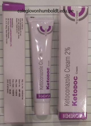
Purchase ketoconazole cream 15gm fast delivery
Disinfectants containing chlorine, iodine, and glutaraldehyde present potent anti� hepatitis B exercise. Culture and Diagnosis and Virulence Factors the hepatitis B virus enters the physique by way of a break in the skin or mucous membrane or by injection into the bloodstream. Eventually, it reaches the liver cells (hepatocytes), the place it multiplies and releases viruses into the blood throughout an incubation period of four to 24 weeks (7 weeks average). Chronic an infection without overt symptoms typically results in a condition called necroinflammation, in which protracted inflammation attributable to the presence of the virus results in liver disease. These exams are important for screening blood destined for transfusions, semen in sperm banks, and organs meant for transplant. Prevention and Treatment and Epidemiology An important factor within the transmission pattern of hepatitis B virus is that it multiplies completely in the liver, which repeatedly seeds the blood with viruses. Electron microscopic research have revealed as a lot as 107 virions per milliliter of infected blood. Even a minute quantity of blood (a millionth of a milliliter) can transmit an infection. The abundance of circulating virions is so high and the minimal dose so low that such easy practices as sharing a toothbrush or a razor can transmit the an infection. Growing concerns about virus unfold through donated organs and tissue are prompting elevated testing previous to surgery. Spread of the virus by the use of shut contact in families or establishments can additionally be nicely documented. Vertical transmission is feasible, and it predisposes the kid to growth of the provider state and elevated danger of liver cancer. The most generally used vaccines are recombinant, containing the pure surface antigen cloned in yeast cells. Vaccination is a must for medical and dental employees and college students, sufferers receiving multiple transfusions, immunodeficient individuals, and cancer sufferers. The vaccine is also now strongly really helpful for all newborns as part of a routine immunization schedule. Another group for whom passive immunization is very really helpful is neonates born to infected moms. Mild instances of hepatitis B are managed by symptomatic therapy and supportive care. Chronic infection can be controlled with recombinant human interferon, tenofovir, or entecavir. Each of these might help to gradual virus multiplication and forestall liver damage in many but not all sufferers. Hepatitis C Virus Hepatitis C is usually referred to as the "silent epidemic" because three. Pregnant women in their third trimester are at highest danger for this severe type of disease, and the fatality rate is nearly 20%. A majority of the instances reported in the United States occur in individuals who have traveled to these endemic regions. Hepatitis E virus is transmitted by the fecal-oral route, mainly by way of contaminated water and food. Pathogenesis and Virulence Factors the virus is so adept at establishing persistent infections that researchers are learning the ways in which it evades immunologic detection and destruction. List the potential causative brokers for the next infectious gastrointestinal circumstances: dental caries, periodontal illnesses, mumps, and gastric ulcers. Differentiate among the major types of hepatitis and discuss causative agents, modes of transmission, diagnostic strategies, prevention, and treatment of each. It is more commonly transmitted by way of blood contact (both "sanctioned," corresponding to in blood transfusions, and "unsanctioned," such as needle sharing by injecting drug users) than by way of transfer of other physique fluids. Anyone with a history of exposure to blood products or organs before 1992 (when effective screening became available) is at larger threat for this an infection, as is anyone with a history of injecting drug use. It has a very excessive prevalence in elements of South America, Central Africa, and China.
Discount ketoconazole cream american express
Here, it anastomoses with the posterior ulnar recurrent and inferior ulnar collateral arteries, participating within the peri-articular arterial anastomoses of the elbow. It then passes inferomedially anterior to the medial epicondyle of the humerus and joins the peri-articular arterial anastomoses of the elbow area by anastomosing with the anterior ulnar recurrent artery. Veins of Arm Two sets of veins of the arm, superficial and deep, anastomose freely with one another. The superficial veins are in the subcutaneous tissue, and the deep veins accompany the arteries. Their frequent connections encompass the artery, forming an anastomotic network inside a typical vascular sheath. The 537 pulsations of the brachial artery help move the blood through this venous community. Not uncommonly, the deep veins join to type one brachial vein throughout part of their course. Their origins from the brachial plexus, courses within the upper limb, and the constructions innervated by them are summarized in Table 3. After crossing the anterior aspect of the elbow, it continues to supply the skin of the lateral facet of the forearm. The branch of the radial nerve to the lateral head of the triceps arises inside the radial groove. Anterior to the lateral epicondyle, the radial nerve divides into deep and superficial branches. The deep department of the radial nerve is completely muscular and articular in its distribution. The superficial branch of the radial nerve is totally cutaneous in its distribution, supplying sensation to the dorsum of the hand and fingers. The median nerve has no branches in the axilla or arm, however it does supply articular branches to the elbow joint. Posterior to the medial epicondyle, the place the ulnar nerve is referred to in lay terms because the "funny bone. Like the median nerve, the ulnar nerve has no branches in the arm, nevertheless it additionally provides articular branches to the elbow joint. Medially, the mass of flexor muscle tissue of the forearm arising from the common flexor attachment on the medial epicondyle; most specifically, the pronator teres. Laterally, the mass of extensor muscles of the forearm arising from the lateral epicondyle and supra-epicondylar ridge; most particularly, the brachioradialis. The floor of the cubital fossa is shaped by the brachialis and supinator muscular tissues of the arm and forearm, respectively. Radial nerve, deep between the muscular tissues forming the lateral boundary of the fossa (the brachioradialis, in particular) and the brachialis, dividing into its superficial and deep branches. Surface Anatomy of Arm and Cubital Fossa the borders of the deltoid are visible when the arm is kidnapped towards resistance. The long, lateral, and medial heads of the triceps brachii type bulges on the posterior aspect of the arm and are identifiable when the forearm is extended from the flexed position towards resistance. It is separated from the pores and skin by only the olecranon bursa, which accounts for the mobility of the overlying pores and skin. The triceps tendon is definitely felt because it descends alongside the posterior aspect of the arm to the olecranon. The fingers could be pressed inward on all sides of the tendon, the place the elbow joint is superficial. The biceps brachii tendon could be palpated in the cubital fossa, instantly lateral to the midline, particularly when the elbow is flexed against resistance. The proximal part of the bicipital aponeurosis could be palpated the place it passes obliquely over the brachial artery and median nerve. The cephalic vein runs superiorly in the lateral bicipital groove, and the basilic vein ascends in the medial bicipital groove. No a part of the shaft of the humerus is subcutaneous; nevertheless, it can be palpated with varying distinctness via the muscles surrounding it, particularly in many elderly individuals. The head of the humerus is surrounded by muscles on all sides, besides inferiorly; thus, it might be palpated by pushing the fingers well up into the axilla. The humeral head could be palpated when the arm is moved whereas the inferior angle of the scapula is held in place.
Diseases
- Hemorrhagic fever with renal syndrome
- Hypercholesterolemia due to LDL receptor deficiency
- Apert like polydactyly syndrome
- Herpes zoster
- Uniparental disomy of 13
- Kozlowski Warren Fisher syndrome
Generic 15gm ketoconazole cream
Describe the method of nitrogen fixation, and explain how microbes play a role in this biochemical response. It is also an necessary habitat for microbes that decompose the advanced litter and gradually recycle nutrients. The humus content varies with local weather, temperature, moisture and mineral content, and microbial motion. For occasion, the moisture and warmth of the tropics promote fast microbial decomposition and thereby cut back humus ranges, and the high ranges of precipitation wash away the nutrients mobilized by the microbes. Native tropical rain forests are adapted to these situations and therefore thrive, whereas agricultural crops do poorly without inputs of fertilizer. On the other hand, the reasonable climate of the temperate zone provides a stability of plant growth and microbial decomposition that causes accumulations of humus, and naturally fertile soils. The biota in soil thrive in cool, dry conditions-quite a special surroundings than the human body. Bacteria which would possibly be able to forming endospores survive in the endospore type for years in soil. These embody the bacteria causing tetanus (Clostridium tetani) and anthrax (Bacillus anthracis). Living Activities in Soil the wealthy tradition medium of the soil supports a incredible array of microorganisms (bacteria, fungi, algae, protozoa, and viruses). These symbiotic associations between fungi and plant roots favor the absorption of water and minerals from the soil. The fungus (darker brown) surrounds the skin of the foundation and penetrates inside it. A typical soil habitat accommodates a mix of clay, silt, and sand together with soil natural matter. Roots and animals (such as, nematodes and mites), in addition to protozoa and micro organism, eat oxygen, which quickly diffuses into the soil pores the place the microbes stay. Note that two kinds of fungi are present: mycorrhizal fungi, which derive their natural carbon from plant roots; and saprophytic fungi, which help degrade natural materials. Some of essentially the most distinctive organic interactions happen within the rhizosphere, the zone of soil surrounding the roots of crops, which accommodates associated micro organism, fungi, and protozoa (see determine 24. Studies have proven that a rich microbial group grows in a biofilm around the root hairs and different exposed surfaces. Their presence stimulates the plant to exude growth components such as carbon dioxide, sugars, amino acids, and vitamins. These vitamins are released into fluid spaces, where they are often readily captured by microbes. Bacteria and fungi likewise contribute to plant survival by releasing hormonelike growth factors and protective substances. We beforehand observed that crops can type close symbiotic associations with microbes to fix nitrogen. Other mutualistic partnerships between plant roots and microbes are mycorrhizae (my-koh-ry-zee). These associations occur when varied species of basidiomycetes, ascomycetes, or zygomycetes attach themselves to the roots of vascular plants (figure 24. The plant feeds the fungus by way of photosynthesis, and the fungus sustains the relationship in a number of ways. By extending its mycelium into the rhizosphere, it helps anchor the plant and increases the floor space for capturing water from dry soils and minerals from poor soils. Plants with mycorrhizae can inhabit extreme habitats more efficiently than vegetation with out them. The topsoil, which extends a few inches to a quantity of ft from the surface, helps a number of burrowing animals similar to nematodes, termites, and earthworms. Aerobic micro organism provoke the digestion of organic matter into carbon dioxide and water and generate minerals such as sulfate, phosphate, and nitrate, which may be further degraded by anaerobic micro organism. Fungal enzymes enhance the effectivity of soil decomposition by hydrolyzing complex pure substances corresponding to cellulose, keratin, lignin, chitin, and paraffin.
Buy ketoconazole cream with paypal
Fetal cardiac results of doxorubicin therapy for carcinoma of the breast throughout pregnancy: case report and evaluate of the literature. Long-term analysis of cardiac perform in kids who received anthracyclines throughout being pregnant. Transplacental switch of anthracyclines, vinblastine, and 4-hydroxycyclophosphamide in a baboon model. Peripartum cardiomyopathy: National Heart, Lung, and Blood Institute and Office of Rare Diseases (National Institutes of Health) workshop suggestions and review. Clinical coronary heart failure during pregnancy and delivery in a cohort of female childhood most cancers survivors treated with anthracyclines. Peripartum cardiomyopathy occurring in a patient beforehand treated with doxorubicin. This is certainly additionally the case for cardiovascular operate and maternal hemodynamics. Gestational complications develop when an organ system is unable to meet the increased physiological calls for of pregnancy. Preeclampsia could also be considered as a derangement of the hemodynamic and cardiovascular system during being pregnant. During later life the hemodynamic and cardiovascular system once more derails when aging takes its toll. Cardiovascular morbidity and mortality is greatly elevated after pregnancies troubled by gestational problems. This is true not only for preeclampsia and other hypertensive issues in being pregnant, but additionally for gestational complications corresponding to preterm supply, gestational diabetes, fetal progress restriction and placental abruption. Excessive weight achieve and weight retention postpartum also pose dangers for maternal health in later life. Gestational problems need to be acknowledged by well being care providers as a danger issue for later cardiovascular disease. The care of women after earlier gestational problems has to be focused on prevention and early detection of indicators of cardiovascular deterioration. Introduction During uncomplicated pregnancies, whole blood volume shows a rise of more than 40%, of which both plasma volume and, to a lesser extent, erythrocyte quantity show giant increments. There is a definite relationship with the relative enhance in complete blood volume and fetal weight. These changes come up roughly concurrently with the modifications in quantity homeostasis but are inclined to precede these changes by some weeks. Gestational issues, and especially the hypertensive syndromes, present an aberrant cardiovascular and hemodynamic adaptation to pregnancy. During otherwise uncomplicated pregnancies there are additionally main adjustments occurring within the metabolic system, such as increased insulin resistance, upregulation of the inflammatory cascade and dyslipidemia. Metabolic changes that resemble adjustments which are seen within the metabolic syndrome are even exaggerated throughout gestational problems. It has lengthy been thought that the consequences of those gestational syndromes are reversed when 229 230 Section 5: Controversies the infant is delivered and (most) values return to normal. However, the detrimental results of the gestational syndromes in combination with the impact of growing older could precipitate those women later in life to develop chronic disease. This means that in later life the effect of aging of the cardiovascular system in these ladies leads to an increased danger of creating cardiovascular morbidity and mortality. The restricted reserves of the organ system of these women at risk shall be unmasked during pregnancy and kind a severe warning. Therefore, being pregnant is the last word stress check for an early failure of organ systems in later life. This may also explain why ladies with pre-existing illness could also be struck extra frequently by gestational issues [1]. History Already during the nineteenth century, German scientists reported that previously preeclamptic women had continual impairment of their renal perform. In that period preeclampsia was still thought of as a renal illness, and that when the illness was maintained long sufficient, renal injury would ensue [2].
15gm ketoconazole cream sale
The child is suddenly lifted (jerked) by the higher limb while the forearm is pronated. The baby may cry out, refuse to use the 708 limb, and protect their limb by holding it with the elbow flexed and the forearm pronated. The proximal a half of the torn ligament could turn into trapped between the pinnacle of the radius and the capitulum of the humerus. The tear within the anular ligament heals when the limb is placed in a sling for 2 weeks. The lunate is pushed out of its place in the flooring of the carpal tunnel toward the palmar floor of the wrist. The displaced lunate might compress the median nerve and result in carpal tunnel syndrome (see scientific box earlier in this chapter). In degenerative joint disease of the wrist, surgical fusion of carpals (arthrodesis) could additionally be essential to relieve the extreme pain. When the 711 epiphysis is placed in its normal position throughout reduction, the prognosis for regular bone progress is good. Elbow joint: Although the elbow joint appears simple due to its primary function as a hinge joint, the fact that it entails the articulation of a single bone proximally with two bones distally, one of which rotates, confers stunning complexity on this compound (three-part) joint. Radio-ulnar joints: the combined synovial proximal and distal radioulnar joints plus the interosseous membrane allow pronation and supination of the forearm. Wrist joint: Motion on the wrist strikes the complete hand, making a dynamic contribution to a ability or motion, or permitting it to be stabilized in a selected place to maximize the effectiveness of the hand and fingers in manipulating and holding objects. The scapulothoracic joint is where the scapular actions of elevation�depression, protraction�retraction, and rotation happen. The scientific community has proposed and widely adopted the use of the extra structurally based mostly time period transverse carpal ligament to replace the time period flexor retinaculum. Commonly, the time period chest is used as a synonym for thorax; nevertheless, the chest is far more in depth than the thoracic wall and cavity contained inside it. The thoracic cavity and its wall have the shape of a truncated cone, being narrowest superiorly, with the circumference growing inferiorly, and reaching its most measurement at the junction with the belly portion of the trunk. The wall of the thoracic cavity is comparatively skinny, basically as thick as its skeleton. Furthermore, the ground of the thoracic cavity (thoracic diaphragm) is deeply invaginated inferiorly. Consequently, almost the lower half of the thoracic wall surrounds and protects stomach rather than thoracic viscera. Thus, the thorax and 718 its cavity are a lot smaller than one would possibly anticipate primarily based on the exterior appearance of the chest. The osteocartilaginous thoracic cage consists of the sternum, 12 pairs of ribs and costal cartilages, and 12 thoracic vertebrae and 719 intervertebral discs. The clavicles and scapulae form the pectoral (shoulder) girdle, one aspect of which is included right here to show the relationship between the thoracic (axial) and upper limb (appendicular) skeletons. The red dotted line indicates the place of the diaphragm, which separates the thoracic and belly cavities. The thorax includes the primary organs of the respiratory and cardiovascular systems. The thoracic cavity is split into three major areas: the central compartment or mediastinum that houses the thoracic viscera apart from the lungs and, on both sides, the best and left pulmonary cavities housing the lungs. The majority of the thoracic cavity is occupied by the lungs, which offer for the trade of oxygen and carbon dioxide between the air and blood. Most of the remainder of the thoracic cavity is occupied by the heart and buildings concerned in conducting the air and blood to and from the lungs. Also, the esophagus, a tubular construction carrying vitamins (food) to the stomach, traverses the thoracic cavity. In phrases of operate and development, the breasts are associated to the reproductive system; nevertheless, the breasts are located on and sometimes dissected with the thoracic wall and therefore are included on this chapter.
Ketoconazole cream 15 gm with mastercard
Specialized lymphatic vessels in the intestinal villi (tiny projections of the mucous membrane) that take up fats are called lacteals. They empty their milklike fluid into the lymphatic plexuses within the walls of the jejunum and ileum. The lacteals drain in turn into lymphatic vessels between the layers of the mesentery. The superior nodes type a system in which the central nodes, at the root of the superior mesenteric artery, obtain lymph from the mesenteric, ileocolic, proper colic, and middle colic nodes, which in turn receive lymph from juxta-intestinal lymph nodes. Efferent lymphatic vessels from the mesenteric lymph nodes drain to the 1103 superior mesenteric lymph nodes. Lymphatic vessels from the terminal ileum observe the ileal department of the ileocolic artery to the ileocolic lymph nodes. The sympathetic fibers within the nerves to the jejunum and ileum originate in the T8�T10 segments of the spinal twine and reach the superior mesenteric nerve plexus by way of the sympathetic trunks and thoracic abdominopelvic (greater, lesser, and least) splanchnic nerves. The presynaptic sympathetic fibers synapse on cell bodies of postsynaptic sympathetic neurons in the celiac and superior mesenteric (prevertebral) ganglia. The parasympathetic fibers in the nerves to the jejunum and ileum derive from the posterior vagal trunks. The presynaptic parasympathetic fibers synapse with postsynaptic parasympathetic neurons within the myenteric and submucosal plexuses of the enteric nervous system in the intestinal wall (see additionally "Summary of Innervation of Abdominal Viscera," p. Presynaptic sympathetic nerve fibers originate in the T8 or T9 by way of T10 or T11 segments of the spinal wire and reach the celiac plexus through the sympathetic trunks and larger and lesser (abdominopelvic) splanchnic nerves. After synapsing in the celiac and superior mesenteric ganglia, postsynaptic nerve fibers accompany the arteries to the gut. Presynaptic parasympathetic (vagus) nerves originate in the medulla (oblongata) and cross to the intestine by way of the posterior 1105 vagal trunk. They synapse with intrinsic postsynaptic neurons of the enteric nervous system positioned within the intestinal wall. Sympathetic stimulation reduces peristaltic and secretory exercise of the intestine and causes vasoconstriction, reducing or stopping gastrointestinal exercise and making blood (and energy) obtainable for "fleeing or fighting. Cessation of sympathetic stimulation permits vasodilation, restoring blood flow to the active bowel. The small gut also has extrinsic and intrinsic sensory (visceral afferent) fibers. Visceral pain from the small intestine could also be referred to dermatomes provided by somatic afferent fibers sharing by the identical spinal sensory ganglia and spinal cord segments. To examine the colon, a barium enema has been given after the bowel is cleared of fecal material by a cleansing enema. Single-contrast barium studies reveal the semilunar folds demarcating the haustra. Following the single-contrast examine, the patient has evacuated the barium and the colon was distended with air for this double-contrast research. Teniae coli: three distinct longitudinal bands: (1) mesocolic tenia, to which the transverse and sigmoid mesocolons connect; (2) omental tenia, to which the omental appendices attach; and (3) free tenia (L. The teniae coli (thickened bands of smooth muscle representing many of the longitudinal coat) begin at the base of the appendix as the thick longitudinal layer of the appendix separates into three bands. The teniae run the length of the massive intestine, abruptly broadening and merging with one another once more at the rectosigmoid junction right into a continuous longitudinal layer around the rectum. If distended with feces or gas, the cecum may be palpable through the anterolateral stomach wall. The frenulum is a fold (more evident in cadavers) that runs from the ileocecal valve alongside the wall on the junction of the cecum and ascending colon. The interior of the cecum showing the endoscopic (living) look of the ileocecal valve. The approximate incidences of various areas of the appendix, primarily based on an evaluation of 10,000 cases, are shown. The terminal ileum enters the 1109 cecum obliquely and partly invaginates into it. It was believed that when the cecum is distended or when it contracts, the lips and frenula actively tighten, closing the valve to forestall reflux from the cecum into the ileum. The round muscle is poorly developed around the orifice; therefore, the valve is unlikely to have any sphincteric motion that controls passage of the intestinal contents from the ileum into the cecum. The papilla most likely serves as a comparatively passive flap valve, stopping reflux from the cecum into the ileum as contractions occur to propel contents up the ascending colon and into the transverse colon (Magee and Dalley, 1986).
Khurasani-Ajavayan (Henbane). Ketoconazole Cream.
- Treating scar tissue, when the leaf oil is used on the skin.
- Are there any interactions with medications?
- Spasms of the digestive tract, including the stomach and intestines.
- Are there safety concerns?
- How does Henbane work?
- What is Henbane?
- Dosing considerations for Henbane.
Source: http://www.rxlist.com/script/main/art.asp?articlekey=96126
Discount ketoconazole cream 15gm without prescription
Ulnar canal syndrome (Guyon tunnel syndrome) is manifest by hypoesthesia (reduced sense of touch or sensation) in the medial one and a half fingers and weak spot of the intrinsic muscles of the hand. This kind of nerve compression, which has been called handlebar neuropathy, leads to sensory loss on the medial facet of the hand and weakness of the intrinsic hand muscle tissue. Radial Nerve Injury in Arm and Hand Disability Although the radial nerve provides no muscular tissues within the hand, radial nerve injury within the arm can produce critical hand incapacity. The hand is flexed at the wrist and lies flaccid, a situation generally known as 646 wrist-drop (see the medical box "Injury to Radial Nerve in Arm"). The fingers of the relaxed hand also stay in the flexed place on the metacarpophalangeal joints. The loss of the flexibility to lengthen the wrist impacts the length rigidity relationship of the wrist and finger flexors. The interphalangeal joints could be prolonged weakly by way of the action of the intact lumbricals and interossei, which are supplied by the median and ulnar nerves (Table 3. Thus, the extent of anesthesia is minimal, even in critical radial nerve accidents, and is usually confined to a small space on the lateral a part of the dorsum of the hand. Dermatoglyphics the science of learning ridge patterns of the palm, called dermatoglyphics, is a priceless extension of the conventional bodily examination of individuals with certain congenital anomalies and genetic illnesses. For instance, individuals with trisomy 21 (Down syndrome) have dermatoglyphics that are extremely attribute. In addition, they often have a single transverse palmar crease (Simian crease); however, roughly 1% of the general population has this crease with no other scientific options of the syndrome. Palmar Wounds and Surgical Incisions the location of superficial and deep palmar arches ought to be kept in mind when examining wounds of the palm and when making palmar incisions. As mentioned beforehand, incisions or wounds along the medial floor of the thenar eminence may injure the recurrent branch of the median nerve to the thenar muscular tissues (see the medical box "Trauma to Median Nerve"). Organization: the muscles and tendons of the hand are organized into 5 fascial compartments: two radial compartments (thenar and adductor) that serve the thumb, an ulnar (hypothenar) compartment that serves the little finger, and two extra central compartments that serve the medial 4 digits (a palmar one for the lengthy flexor tendons and lumbricals, and a deep one between the metacarpals for the interossei). Muscles: the greatest mass of intrinsic muscular tissues is dedicated to the extremely cellular thumb. Indeed, when extrinsic muscles are additionally thought of, the thumb has eight muscles producing and controlling the big range of actions that distinguish the human hand. Vasculature: the vasculature of the hand is characterized by multiple anastomoses between both radial and ulnar vessels and palmar and dorsal vessels. Thus, blood is generally out there to all parts of the hand in all positions as nicely as whereas performing features (gripping or pressing) which may in any other case compromise particularly the palmar structures. Innervation: Unlike the dermatomes of the trunk and proximal limbs, the zones of cutaneous innervation and the roles of motor innervation are properly 648 outlined, as are functional deficits. The clavicle varieties a strut (extension) that holds the scapula, therefore the glenohumeral (shoulder) joint, away from the thorax so it could possibly move freely. The pectoral girdle is a partial bony ring (incomplete posteriorly) fashioned by the manubrium of the sternum, the clavicle, and the scapulae. Joints related to these bones are the sternoclavicular, acromioclavicular, and glenohumeral. The girdle offers for attachment of the superior appendicular skeleton to the axial skeleton and offers the mobile base from which the higher limb operates. When testing the vary of movement of the pectoral girdle, each scapulothoracic (movement of the scapula on the thoracic wall) and glenohumeral actions must be thought of. Although the initial 30� of abduction might occur without scapular motion, in the general movement of totally elevating the arm, the movement occurs in a 2:1 ratio: For each 3� of elevation, approximately 2� happens on the glenohumeral joint and 1� on the physiological scapulothoracic joint. Hence, when the upper limb has been elevated so that the arm is vertical at the facet of the top (180� of arm abduction or flexion), 120� occurred on the glenohumeral joint and 60� occurred on the scapulothoracic joint. Thus, though the articular disc serves as a shock absorber of forces transmitted alongside the clavicle from the upper limb, dislocation of the clavicle is uncommon, whereas fracture of the clavicle is widespread. It is hooked up to the margins of the articular surfaces, including the periphery of the articular disc. A synovial membrane lines the internal floor of the fibrous layer of the joint capsule, extending to the perimeters of the articular surfaces. Anterior and posterior sternoclavicular ligaments reinforce the joint capsule anteriorly and posteriorly. It extends from the sternal end of 1 clavicle to the sternal end of the other clavicle.
Buy ketoconazole cream 15 gm overnight delivery
The last effects rely upon the kinds and numbers of microbes and whether the meals is cooked or preserved. In some circumstances, specific microbes can even be added to food to get hold of a desired effect. The results of microorganisms Bread Microorganisms accomplish three functions in bread making: 1. Leavening is achieved primarily by way of the release of fuel to produce a porous and spongy product. Other gasforming microbes such as coliform bacteria, certain Clostridium 760 Chapter 25 Applied Microbiology and Food and Water Safety species, heterofermentative lactic acid bacteria, and wild yeasts could be employed, depending on the sort of bread desired. Yeast metabolism requires a supply of fermentable sugar such as maltose or glucose. Because the yeast respires aerobically in bread dough, the chief merchandise of maltose fermentation are carbon dioxide and water, somewhat than alcohol (the primary product in beer and wine). Other contributions to bread texture come from kneading, which contains air into the dough, and from microbial enzymes, which break down flour proteins (gluten) and provides the dough elasticity. Besides carbon dioxide production, bread fermentation generates different volatile natural acids and alcohols that impart delicate flavors and aromas. These are particularly well-developed in handmade bread, which is leavened extra slowly than industrial bread. Yeasts and micro organism can even impart unique flavors, depending upon the culture mixture and baking methods used. The pungent flavor of rye bread, for instance, is available in half from starter cultures of lactic acid bacteria such as Lactobacillus plantarum, L. The starting ingredients for both ancient and present-day versions of beer, ale, stout, porter, and other variations are water, malt (barley grain), hops, and special strains of yeasts. The steps in brewing embody malting, mashing, adding hops, fermenting, aging, and finishing. This process, known as malting, releases amylases that convert starch to dextrins and maltose, and proteases that digest proteins. Other sugar and starch supplements added in some forms of beer are corn, rice, wheat, soybeans, potatoes, and sorghum. After the sprouts have been separated, the remaining malt grain is dried and saved in preparation for mashing. Sugar and starch supplements are then launched to the mash mixture, which is heated to a temperature of about 65�C to 70�C. During this step, the starch is hydrolyzed by amylase and simple sugars are launched. Boiling additionally caramelizes the sugar and imparts a golden or brown colour, destroys any bacterial contaminants that can destroy taste, and concentrates the mixture. The filtered and cooled supernatant is then ready for the addition of yeasts and fermentation. Fermentation begins when wort is inoculated with a species of Saccharomyces that has been specifically developed for beer making. Top yeasts corresponding to Saccharomyces cerevisiae perform at the surface and are used to produce the higher alcohol content of ales. In each instances, the preliminary inoculum of yeast starter is aerated briefly to promote fast progress and improve the load of yeast cells. Shortly thereafter, an insulating blanket of foam and carbon dioxide develops on the surface of the vat and promotes anaerobic situations (figure 25. During 8 to 14 days of fermentation, the wort sugar Anaerobic circumstances in do-it-yourself beer production. The variety of flavors within the completed product is partly as a result of the release of small quantities of glycerol, acetic acid, and esters. Fermentation is self-limited, and it primarily ceases when a concentration of 3% to 6% ethyl alcohol is reached. During this maturation interval, yeast, proteins, resin, and different supplies settle, forsaking a clear, mellow fluid. Lager beer is subjected to a final filtration step to remove any residual yeasts that would spoil it. Some winemakers enable these natural yeasts to dominate, however many wineries inoculate the must with a particular strain of Saccharomyces cerevisiae, variety ellipsoideus. To discourage yeast and bacterial spoilage brokers, winemakers sometimes deal with grapes with sulfur dioxide or potassium metabisulfite.
Generic ketoconazole cream 15gm fast delivery
Roof: formed laterally by the transversalis fascia, centrally by musculoaponeurotic arches of the internal indirect and transversus abdominis, and medially by the medial crus of the external oblique aponeurosis. Floor: fashioned laterally by the iliopubic tract, centrally by gutter formed by the infolded inguinal ligament, and medially by the lacunar ligament. The inguinal triangle separates these formations from the constructions of the femoral sheath (femoral vessels and femoral canal) that traverse the medial part of the subinguinal space. Most groin hernias in males pass superior to the iliopubic tract (inguinal hernias), whereas most pass inferior to it in females (femoral hernias). Because of its relative weak point, the myopectineal orifice is overlaid with prosthetic mesh positioned in the extraperitoneal retro-inguinal area ("area of Bogros") in lots of hernia repairs. The male gubernaculum is a fibrous tract connecting the primordial testis to the anterolateral stomach wall at the web site of the longer term deep ring of the inguinal canal. A peritoneal diverticulum, the processus vaginalis, traverses the creating inguinal canal, carrying muscular and fascial layers of the anterolateral abdominal wall before it because it enters the primordial scrotum. The testis begins to cross by way of the inguinal canal during the twenty eighth week and takes approximately three days to traverse it. The stalk of the processus vaginalis normally degenerates; nonetheless, its distal saccular half forms the tunica vaginalis, the serous sheath of the testis and epididymis (Moore et al. A fetus at 28 weeks (7th month) reveals the processus vaginalis and testis passing via the inguinal canal. In a new child toddler, obliteration of the stalk of the processus vaginalis has occurred. The remains of the processus vaginalis have formed the tunica vaginalis of the testis. The processus vaginalis of the peritoneum traverses the transversalis fascia on the website of the deep inguinal ring, forming the inguinal canal as within the male, and protrudes into the creating labium majus, which is the feminine homologue of (part corresponding to) the scrotum. At 2 months, the undifferentiated gonads (primordial ovaries) are situated on the dorsal belly wall. The processus vaginalis (not shown) passes via the abdominal wall, forming the inguinal canal on all sides as in the male fetus. The round ligament passes via the canal and attaches to the subcutaneous tissue of the labium majus. In the mature female, the processus vaginalis has degenerated, but the round ligament persists and passes via the inguinal canal. The female gubernaculum, a fibrous twine connecting the ovary and primordial uterus to the creating labium majus, is represented postnatally by the ovarian ligament, between the ovary and uterus, and the round ligament of the uterus (L. Except for its most inferior half, which turns into a serous sac engulfing the testis, the tunica vaginalis, the processus vaginalis obliterates by the sixth month of fetal improvement. The inguinal canals in females are narrower than those in males, and the canals in infants of each sexes are shorter and far less oblique than in adults. The superficial inguinal rings in infants lie almost instantly anterior to the deep inguinal rings. Consequently, increases in intra-abdominal stress act on the inguinal canal, forcing the posterior wall of the canal against the anterior wall and strengthening this wall, thereby decreasing the probability of herniation till the pressures overcome the resistant effect of this mechanism. Simultaneously, contraction of the external oblique approximates the anterior wall of the canal to the posterior wall. It additionally increases pressure on the medial and lateral crura, resisting enlargement (dilation) of the superficial inguinal ring. The inguinal canal consists of a series of three musculo-aponeurotic arcades traversed by the spermatic cord or round ligament of the uterus (arrow). The spermatic cord begins on the deep inguinal ring lateral to the inferior epigastric vessels, passes through the inguinal canal, exits on the superficial inguinal ring, and ends within the scrotum at the posterior border of the testis. Fascial coverings derived from the anterolateral stomach wall during prenatal development encompass the spermatic twine. Arterial supply and lymphatic drainage of the testis and scrotum; innervation of the scrotum. The lumbar plexus offers innervation to the anterolateral aspect of the scrotum; the sacral plexus provides innervation to the postero-inferior side. Cremasteric fascia: derived from the investing fascia of each the superficial and deep surfaces of the interior indirect muscle. External spermatic fascia: derived from the exterior indirect aponeurosis and its investing fascia. The cremaster muscle reflexively attracts the testis superiorly within the scrotum, particularly in response to chilly.
Order cheap ketoconazole cream line
Two pulmonary veins, a superior and an inferior pulmonary vein on both sides, carry oxygen-rich ("arterial") blood from corresponding lobes of every lung to the left atrium of the center. Except in the central, perihilar area of the lung, the veins from the visceral pleura and the bronchial venous circulation drain into the pulmonary veins, the comparatively small quantity of low-oxygen blood getting into the massive quantity of oxygen-rich blood returning to the guts. Veins from the parietal pleura join systemic veins in adjacent elements of the thoracic wall. The single right bronchial artery can also arise instantly from the aorta; nevertheless, it commonly arises not directly, both by way of the proximal a part of one of the higher posterior intercostal arteries (usually the proper third posterior intercostal artery) or from a typical trunk with the left superior bronchial artery. The bronchial arteries provide the supporting tissues of the lungs and visceral pleura. The bronchial veins drain the more proximal capillary beds supplied by the bronchial arteries; the remaining is drained by the pulmonary veins. Then, they usually move alongside the posterior elements of the principle bronchi, supplying them and their branches as far distally as the respiratory bronchioles. The the rest of the blood is drained by the pulmonary veins, particularly the blood coming back from the visceral pleura, the extra peripheral regions of the lung, and the distal parts of the foundation of the lung. The proper bronchial vein drains into the azygos vein, and the left bronchial vein drains into the accent hemi-azygos vein or the left superior intercostal vein. The superficial subpleural lymphatic plexus lies deep to the visceral pleura and drains the lung parenchyma (tissue) and visceral pleura. Lymphatic vessels from this superficial plexus drain into the bronchopulmonary lymph nodes (hilar lymph nodes) in the region of the lung hilum. The lymphatic vessels originate from superficial subpleural and deep lymphatic plexuses. All lymph from the lung leaves alongside the basis of the lung and drains to the inferior or superior tracheobronchial lymph nodes. The inferior lobe of each lungs drains to the centrally placed inferior tracheobronchial (carinal) nodes, which primarily drain to the best facet. The different lobes of every lung drain primarily to the ipsilateral superior tracheobronchial lymph nodes. From right here, the lymph traverses a variable number of paratracheal nodes and enters the bronchomediastinal trunks. The deep bronchopulmonary lymphatic plexus is positioned within the submucosa of the bronchi and in the peribronchial connective tissue. It is basically involved with draining the buildings that type the foundation of the lung. Lymphatic vessels from this deep plexus drain initially into the intrinsic pulmonary lymph nodes, positioned along the lobar bronchi. Lymphatic vessels from these nodes proceed to comply with the bronchi and pulmonary vessels to the hilum of the lung, where they also drain into the bronchopulmonary lymph nodes. From them, lymph from 816 each the superficial and deep lymphatic plexuses drains to the superior and inferior tracheobronchial lymph nodes, superior and inferior to the bifurcation of the trachea and major bronchi, respectively. The proper lung drains primarily by way of the consecutive sets of nodes on the proper side, and the superior lobe of the left lung drains primarily by way of corresponding nodes of the left facet. Many, however not all, of the lymphatics from the inferior lobe of the left lung, nevertheless, drain to the right superior tracheobronchial nodes; the lymph then continues to observe the right-side pathway. Lymph from the tracheobronchial lymph nodes passes to the proper and left bronchomediastinal lymph trunks, the main lymph conduits draining the thoracic viscera. These trunks often terminate on both sides on the venous angles (junctions of the subclavian and inner jugular veins); nevertheless, the proper bronchomediastinal trunk might first merge with other lymphatic trunks, converging right here to form the quick right lymphatic duct. Lymph from the parietal pleura drains into the lymph nodes of the thoracic wall (intercostal, parasternal, mediastinal, and phrenic). A few lymphatic vessels from the cervical parietal pleura drain into the axillary lymph nodes. These nerve networks include parasympathetic, sympathetic, and visceral afferent fibers. After contributing to the posterior pulmonary plexus, the vagus nerves proceed inferiorly and become part of the esophageal plexus, typically losing their identification after which reforming as anterior and posterior vagal 818 trunks.
References
- Keszler M, Klein R, McClellan L, et al: Effects of conventional and high frequency jet ventilation on lung parenchyma. Crit Care Med 10:514, 1982.
- Menarini M, Del Popolo G, Di Benedetto P, et al: Trospium chloride in patients with neurogenic detrusor overactivity: is dose titration of benefit to the patients?, Int J Clin Pharmacol Ther 44(12):623, 2006.
- Young DF, Tsai FY: Flow characteristics in models of arterial stenoses. II. Unsteady flow, J Biomech 6:547-559, 1973.
- Horvath KA, Cohn LH, Cooley DA, et al: Transmyocardial laser revascularization: Results of a multicenter trial with transmyocardial laser revascularization used as sole therapy for end-stage coronary artery disease. J Thorac Cardiovasc Surg 1997;113: 645-654.


