Vasotec
Vasotec dosages: 10 mg, 5 mg
Vasotec packs: 60 pills, 90 pills, 120 pills, 180 pills, 270 pills, 360 pills
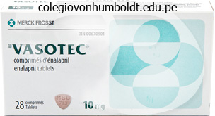
Order vasotec online pills
If enhancements in cranial vault form are to be achieved, most circumstances require a proper complete cranial vault reshaping on the age of four to 8 months. In our heart, a latest trend towards earlier surgical procedure (4 mo) involving elimination of the stenosed suture together with extensive barrel-staving and postoperative helmet-molding is gaining popularity. Early outcomes using this protocol have proven promising outcomes with much less blood loss and shorter operative instances. Variations in the degree of the scaphocephalic deformity are frequent, relying on the extent of sagittal suture stenosis. When the posterior half is fused, the affected person is handled in the inclined position with the posterior two thirds of the cranial vault reshaped. When the anterior half is fused, the affected person is handled in the supine place with the anterior two thirds of the cranial vault reshaped, with or without superior orbital rim reshaping. When the complete suture is fused, a mix of each approaches could additionally be needed. Unless a big concomitant supraorbital deformity exists, we favor to deal with full sagittal suture stenosis (anterior and posterior) at one operative setting in the inclined position via a total cranial vault reshaping. For older youngsters (>1 yr) or kids with a necessity for upper orbital reconstruction, we prefer the supine place at one operative setting or, hardly ever, in two phases, with posterior reconstruction previous anterior and orbital reconstruction by 4 to 6 months. Other centers have reported good results when routinely staging full sagittal synostosis. Unilateral Lambdoid Synostosis Many surgeons contemplate easy strip craniectomy of the involved suture or partial craniectomy of the region to be enough therapy. If enhancements in cranial vault form are required after 10 to 12 months of age, formal posterior cranial vault reshaping is performed. Surgical administration of these sufferers has been advocated to happen from the first few weeks after start until nicely in to the kids. Many of these sufferers require a number of, staged procedures that contain movements of the bone and soft tissue from each the intracranial and the extracranial approaches. The surgical method to most of those congenital deformities was radically modified by strategies launched to the United States by Paul Tessier of France in 1967. From his imaginative intracranial and extracranial approaches, quite a few advances have been made that have improved the administration of these advanced pediatric craniofacial deformities. A 6-month-old woman with anterior and posterior sagittal suture synostosis leading to scaphocephaly. She underwent whole cranial vault reshaping with out the necessity for any orbital osteotomies. G, Prone positioning is important and requires cautious safety of each the airway and the globes. H, Intraoperative superior view of proposed osteotomy sites for whole cranial vault reshaping. I, Intraoperative superior view of the osteotomies, reshaping, and resorbable plate fixation. K, Intraoperative left lateral view after osteotomies, reshaping, and resorbable plate fixation. Perspectives on craniosynostosis: sutural biology, some well-known syndromes, and a few unusual syndromes. Long-term neuropsychologic effects of sagittal craniosynostosis on youngster growth. Intracranial volume and cephalic index outcomes for total calvarial reconstruction among nonsyndromic sagittal synostosis sufferers. Papilledema in isolated single-suture craniosynostosis: prevalence and predictive components. Long-term neuropsychological development in single-suture craniosynostosis handled early. Computer-assisted imaging in the analysis, administration and study of dysmorphic sufferers. Computerized imaging for delicate tissue and osseous reconstruction within the head and neck. In Proceedings of the 6th International Congress on Cleft Palate and Related Craniofacial Anomalies. The early versus the late reconstruction of congenital hypoplasia of the facial skeleton and cranium.
Purchase vasotec overnight
Whereas most maxillectomy defects are best reconstructed primarily, prosthetic obturation of the defect might supply benefits in some conditions, together with an easier operation, easier early detection of recurrence, and substitute of enamel. Dental implants normally, and the zygomatic implant in particular, can provide an unlimited enchancment in prosthetic function. McGuirt and colleagues130 have demonstrated improved consequence with elective remedy of the neck, and elective treatment of the neck must be thought-about in all but the smallest thin lesions (<3 mm). Sagittal, or "inner-table" mandibulectomy may be thought-about in smaller tumors that abut the mandible with out evidence of invasion (see later discussion), but giant inner-table resection is technically tough and can considerably weaken the mandible. Small main cancers can be equally treated with surgical procedure or radiation, though surgery is the selection of most clinicians. Squamous cell carcinoma of the proper soft palate invading posteriorly to contain the anterior pole of the tonsil. Le Fort I island approach to a posterior maxillary tumor, without Weber-Fergusson incision. If superior extension ends in exposure of dura mater as a half of a skull base resection, flap coverage is advisable. Overall 5-year survival for alveolar ridge carcinoma is 50% to 65% for both the upper and the lower alveolar ridge. Poor outcome is associated with superior stage, perineural spread, and constructive margins. In these instances, in addition to nodal involvement, adjunctive radiation is really helpful. This and other reports call in to query the knowledge of remark alone for the clinically negative neck in maxillary and hard palate cancers. The morbidity of this strategy was thought essential to eradicate in-transit metastases, a belief that was likely based on mistranslation by McGregor139 of an article published by Polya and von Navratil in 1902, during which they actually recommended elimination of the periosteum or rim of the mandible and not a section. It was found that squamous cell carcinoma invasion occurs mostly via the periodontal ligament within the dentate mandible and thru the porous occlusal surface of the edentulous mandible. Once the cortex was invaded, the inferior alveolar canal was often involved, especially in edentulous mandibles. It is Palate Squamous cell cancers of the palate, onerous and soft, are rare within the United States, however are more widespread in India owing to the phenomenon of "reverse smoking," which assaults the palate with both carcinogens and warmth. Cancers arising from the exhausting palate, nonetheless, may prolong on to the soft palate and vice versa. The periosteum of the palate acts as a significant barrier, and smaller lesions may be treated with extensive native excision. The affected person should be informed that regardless of preservation of palatal bone, a small fistula should develop through the therapeutic course of. Oral-nasal communications within the onerous palate may be treated with an obturator or a neighborhood flap, corresponding to an anteriorly based mostly midline tongue flap. Oral-nasal fistulas in the taste bud are best treated with momentary obturation, as a end result of the majority will close spontaneously. Previously, it was thought that occult cervical metastases are uncommon amongst maxillary and exhausting palate cancers (10�25%), and elective remedy of the neck was usually not indicated besides in T3 or T4 lesions. The scientific and radiographic evaluations of mandibular involvement are frequently inaccurate. Clinical findings such as impairment of inferior alveolar nerve operate or fixation of the tumor to the mandible increase the index of suspicion. The history of an extraction of a tooth in an space of a cancer could suggest native mandibular invasion. Although used by some, bone scans are cumbersome to get hold of and tough to interpret accurately. A high-quality panoramic radiograph is probably probably the most generally used device to decide on mandibular marginal resection versus segmental resection. Also, newer techniques that modulate the magnetic subject in an try and look at adjustments within the bone marrow maintain promise for evaluating mandibular involvement. Tumor invasion in to the marrow house is accompanied by a decrease intermediate signal. A robust shiny marrow signal associated with normal marrow fats underlying the dark cortex usually excludes mandibular involvement. Some surgeons ship periosteum for frozen-section analysis, whereas others submit cancellous scrapings for frozen evaluation.
Diseases
- Olmsted syndrome
- Atelosteogenesis, type II
- Holmes Collins syndrome
- Metaphyseal anadysplasia
- Deletion 6q16 q21
- Hairy nose tip
- Neuraminidase beta-galactosidase deficiency
Discount vasotec 5 mg otc
Oxygen administered through the endotracheal tube ought to be elevated to an FiO2 of 100% (especially if the affected person is comatose) till arterial blood fuel measurements affirm hemoglobin saturation (PaN 2 > 60�70 mmHg), at which level FiO2 may be lowered to between 40% and 60%. Some concern exists concerning the suppression of the respiratory drive with oxygen remedy, however the hypoxic drive could be reestablished after stabilization of the injured patient. The most necessary mechanism of supply of oxygen to the tissues is the hemoglobin throughout the erythrocytes within the cardiovascular system. In a traumatized patient, hemorrhage Circulation Following establishment of an sufficient airway and respiration in the injured patient, the cardiovascular system of the patient should be assessed and control of baseline circulation to the tissues must be rapidly restored. The most typical reason for shock in the traumatized affected person is hypovolemia attributable to hemorrhage, either externally or internally in to physique cavities. Assessment of the diploma of shock is necessary as a outcome of insufficient tissue perfusion can cause irreversible harm to important organs such as the mind or kidneys in a short while period. During the first evaluation, a minimal of two large-bore (14�16 gauge) intravenous catheters ought to be placed peripherally if fluid resuscitation is required. At the time of placement of an intravenous catheter, blood must be drawn from the catheter to enable for typing, cross-matching, and baseline hematologic and chemical studies. Tissue perfusion and oxygenation are dependent on cardiac output and are greatest initially evaluated by physical examination of skin perfusion, pulse price, and psychological standing of the patient. Blood stress levels are commonly used as a surrogate for cardiac output and to suspect hypovolemia, but within the emergency scenario, blood stress measurement could also be an unreliable indicator of growing shock. The response of the blood stress stage to intravascular loss is nonlinear because compensatory mechanisms of increased cardiac fee and contractility, along with venous and arteriolar vasoconstriction, preserve the blood stress within the young wholesome grownup through the first 15% to 20% of intravascular blood loss. Skin perfusion is essentially the most dependable indicator of poor tissue perfusion in the course of the initial analysis of the patient. The early physiologic compensation for volume loss is vasoconstriction of the vessels to the skin and muscles. The cutaneous capillary beds are one of the first areas to shut down in response to hypovolemia due to stimulus from the sympathetic nervous system and the adrenal gland via epinephrine and norepinephrine launch. The release of the catecholamines causes sweating, and through palpation, the skin may feel cool and damp. The decrease extremities are usually first to be affected, and the primary indication of intravascular loss could additionally be paleness and coolness of the skin over the feet and kneecaps. A examine of the capillary filling time by performing a blanch take a look at offers an estimate of the quantity of blood flowing to the capillary beds. In this check, stress is placed on the fingernail, toenail, or hypothenar eminence of the hand ( to evacuate blood from the capillary beds), adopted by a quick launch of the strain. The time required for the blood to return to the capillary beds, represented by the restoration of normal tissue shade, is normally less than 2 seconds in the normovolemic affected person. However, in adults with tachycardia larger than a hundred and twenty beats per minute (bpm), hypovolemia must be anticipated and investigated additional. Older patients usually are unable to exceed charges of one hundred forty bpm in a hypovolemic state, whereas youthful sufferers may current charges of one hundred sixty to 180 bpm with severe intravascular loss. The most distal palpable pulse could give some indication of the blood pressure and cardiac output. Pulse rhythm and regularity may present clues to increasing hypovolemia and cardiac hypoxia. Cardiac dysrhythmias similar to untimely ventricular contractions or arterial fibrillation produce an irregular price and rhythm, signaling the potential lack of compensating mechanisms maintaining myocardial oxygenation. Decreased intravascular quantity is straight away mirrored in decreased urinary output because the compensatory mechanisms of the physique decrease blood circulate to the kidneys in favor of blood circulate to the guts and brain. Any patient with significant trauma should at all times have an indwelling urinary catheter inserted to monitor urine quantity every 15 minutes. If urethral damage is unlikely, the urinary catheter may be placed with minimal concern after a rectal examination. Classic signs of urethral damage embrace blood on the meatus, scrotal hematoma, or a high-riding boggy prostate on rectal examination. Alterations within the mental status of the trauma affected person caused solely by hypovolemia are unusual, except in essentially the most progressive preterminal stages of intravascular fluid loss. The mental changes normally seen are agitation, confusion, uncooperativeness, anxiousness, and irrationality.
Buy vasotec 5 mg visa
The associated osteomas are often discovered in the jaws, notably in the angle region of the mandible, in addition to the facial bones and long bones. Investigation should embody an in depth history of gastrointestinal disturbance and, if optimistic, follow-up with colonoscopy; if the analysis is confirmed, a prophylactic colectomy is usually recommended. Histologically, they encompass both dense compact bone with sparse marrow areas or lamellar trabeculae of cancellous bone with fibrofatty marrow areas. Panoramic radiograph shows numerous radiopaque overseas our bodies in the proper temporomandibular joint (arrow). Treatment is symptomatic; when symptoms occur, localized excision is really helpful through the traditional temporomandibular strategy. A desmoplastic fibroma presents as a welldefined radiolucency at the decrease border of the body of the left mandible in a 3-year-old affected person. The medical history, immunohistochemistry, or genetic markers should be used to differentiate the lesions. Central Giant Cell Granuloma Central large cell granuloma is a lesion occurring virtually solely in the jaws. It happens primarily in the anterior components of the jaws in individuals in the second and third many years of life, however it has been recorded in all websites at all ages. When first described it was referred to as a "reparative giant cell granuloma,"87�89 and it was thought-about a reparative lesion that was basically self-healing. There was little proof of this, nevertheless, and solely oblique references to its self-healing properties could be found. Most appear to observe a fairly benign course, but more aggressive lesions have been noted. It has been suggested that it could be an inflammatory lesion, a reactive lesion, a real tumor, or an endocrine lesion. A desmoplastic fibroma presents as an illdefined radiolucency of the left mandible causing displacement of teeth in a patient aged 8 years. In treating this lesion, the adage "treat the biology, not the histology" is of paramount importance. This is psychologically tough for the surgeon to perform in a younger youngster without a histologic prognosis of malignancy, however the recurrence fee may be very high after more conservative procedures. For lesions in inaccessible areas corresponding to the base of the cranium, radiation remedy and/or chemotherapy has been attempted with variable levels of success. A central giant cell granuloma of the anterior mandible causing the displacement of tooth. Intralesional steroids (usually triamcinolone injected in to the lesion once per week for 6 wk) have been advocated and have proven some success. A central giant cell granuloma of the left angle area of the mandible, appearing as an ill-defined multilocular radiolucency, causing resorption of the distal root of the primary molar (unusual). Radiographically, the central giant cell granuloma can take numerous types from a well-defined radiolucency, a extra ill-defined radiolucency, or a multilocular radiolucency. Histologically, these granulomas include focal preparations of large cells within a vascular stroma with thinwalled capillaries adjoining to the giant cells. Immunohistochemistry has shown that the enormous cells are actually osteoclasts,ninety four and the spindle cells are probably the cells of origin of this lesion. With the aggressive variants, extra aggressive surgery has been suggested including mandibular resection and appropriate reconstruction. A, A central big cell granuloma of the best mandibular bicuspid region inflicting displacement of the foundation of the first bicuspid (arrow). B, One yr after a course of six intralesional injections of triamcinolone (10 mg/mL). However, in any explicit case, it might be extremely troublesome to make this distinction. Mobilization from bone takes place focally and produces lesions in the bones (including the jaws) that are often identified as brown tumors due to their pretty distinctive coloration on surgical exploration. Therefore, whenever a lesion such as this is recurrent, aggressive, or a quantity of, hyperparathyroidism have to be excluded by means of serum calcium, phosphate, and parathormone and parathormone-related protein assays.
Discount 10 mg vasotec otc
Intraoperative lymphatic mapping and selective cervical lymphadenectomy for early stage melanomas of the top and neck. The use of intraoperative radiolymphoscintigraphy for sentinel node biopsy in patients with malignant melanoma. A new approach to pre-treatment assessment of the N0 neck in oral squamous cell one hundred fifty five. Predictive value of tumor thickness in squamous cell carcinoma confined to the tongue and flooring of the mouth. Tumour thickness predicts cervical nodal metastases and survival in early tongue most cancers. Ultrasonography-guided nice needle aspiration for the assessment of cervical metastases. Positron Eemission tomography within the administration of unknown major head and neck carcinoma. Application of new imaging techniques for the analysis of squamous cell carcinoma of the top and neck. Excision of most cancers of the head and neck with special reference to the plan of dissection primarily based upon one-hundred thirty-two operations. Sentinel lymph node biopsy accurately stages the regional lymph nodes for T1-T2 oral squamous cell carcinomas: outcomes of a potential multi-institutional trial. Who deserves a neck dissection after definitive chemoradiotherapy for N2�N3 squamous cell head and neck cancer N2 illness in patients with head and neck squamous cell most cancers handled with chemoradiotherapy: is there a task for post-treatment neck dissection Supraomohyoid neck dissection within the treatment of head and neck tumors: survival ends in 212 cases. Failure at the major site following multi-modality treatment in advanced head and neck most cancers. Failure at distant websites following multi-modality remedy in advanced head and neck most cancers. Second primary neoplasms in patients successfully handled with multimodality therapy for superior head and neck most cancers. Salvage surgery for sufferers with recurrent squamous cell carcinoma of the aerodigestive tract: when do the ends justify the means Routine long-term follow-up in, sufferers treated with curative intent for squamous cell carcinoma of the larynx, pharynx and oral cavity. Early prediction of response to chemoradiotherpy for head and neck cancer: reliability of restaging with combined positron emission tomography and computed tomography. The position of yearly chest radiography in the early detection of lung cancer following oral most cancers. A half-yearly chest radiograph for early detection of lung cancer following oral most cancers. Controversies in multi-modality remedy for head and neck cancer: scientific and biologic perspectives. Full-dose reirradiation for unresectable head and neck carcinoma: expertise on the Gustave-Roussy Institute in a sequence of 169 sufferers. Phase I trial of mixed chemotherapy and reirradiation for recurrent unresectable head and neck cancer. Concomitant chemotherapy and reirradiation as administration for recurrent most cancers of the head and neck. Prognsotic elements in salvage surgical procedure for recurrent oral and oropharyngeal most cancers. Markers of radioresistance in squamous cell carcinomas of the top and neck: a clinicopathologic and immunohistochemical examine. Perioperative problems, comorbidities, and survival in oral and oropharyngeal cancer. Presentation, therapy and end result of oral cavity cancer: a nationwide most cancers knowledge base report. In the United States, roughly 4300 new circumstances resulting in almost 150 deaths are diagnosed yearly. In some people, lip cancer might behave aggressively, manifested by recurrence or mortality in as much as 15% of patients. The lower lip is the most typical website for lip most cancers (88�98%), with only 2% to 7% arising from the higher lip and 2% to 4% at the oral commissures.
Syndromes
- Thoughts that "jump" between different topics ("loose associations")
- Your back pain extends to the buttocks, arms, or legs
- Constipation
- Gout medicines
- Diabetes
- Myopia
- Permanent deformity
- Bone deformities in the face
- Webbing or fusion of the toes
Purchase vasotec 5mg free shipping
Approximation is the act of bringing the nerve stumps in to direct contact and assessing the degree of Neurorrhaphy Neurorrhaphy is the act of bringing the nerve ends collectively and maintaining this position with sutures. A direct neurorrhaphy is performed with the proximal and distal nerve stumps, and an oblique neurorrhaphy is performed when utilizing an interpositional graft between the nerve stumps, such as a nerve graft or conduit. Size of Donor Nerve Grafts Relative to Injured Nerve Donor Nerve Injured Nerve Inferior alveolar (2. Anatomic-histologic survey of the sural nerve: implications for inferior alveolar nerve grafting. Generally, 8-0 to 10-0 monofilament nonresorbable nylon suture is most popular, because a resorbable suture material would are probably to invoke an inflammatory response and disturb the area of anticipated neural healing. At least two sutures ought to be used per anastomosis site to forestall rotation of the nerve stumps, however no extra than three or four sutures should be used per anastomosis. The second suture is positioned one hundred eighty levels from the first suture row on the lateral side of the repair website, and then an assessment is made regarding the need for the position of one or two further sutures. Sural grafts up to 20 cm in size are possible, and patients tolerate the donor website deficit well. As a department of the cervical plexus (C1, C2), the larger auricular nerve supplies sensation to the pre- and postauricular areas, the lower third of the ear, and the skin overlying the posteroinferior border on the angle of the mandible. Patients are usually not amenable to sacrificing sensation of 1 facial area to regain sensation in one other neighboring location. The sole advantage of a higher auricular graft over a sural graft is in situations when it may be harvested through the identical incision for an additional process, such because the repair of an extraoral mandibular fracture or administration of pathology. The basic premise with graft restore is that the nerve graft will provide the Schwann cell conduit sheaths and the expansion elements essential to support and encourage axonal sprouting by way of the graft towards the goal website. This allograft is avialble in varied lengths and diameters that can be used for the trigeminal system. Although the allograft is decellularized, it mainatins sure neurotrophic and neurotropic elements similar to basement membrane laminin. It ought to be remembered that a poor end result after tried microneurosurgery might preclude future surgical choices; subsequently, the most effective likelihood for microneurosurgical success happens on the first (and more than likely, last) surgical intervention. In basic, the nerve regeneration process progresses at approximately 1 mm/day (~3 cm/mo) from the cell body to the target site. With graft or conduit oblique repair, the timeframe is lengthened owing to slowed regeneration via the graft web site, and restoration is variable. All nerve accidents should be documented and evaluated with a historical past, examination, and neurosensory testing (objective and subjective). In cases of observed or known nerve harm, immediate referral for microsurgery provides the best alternative for sensory recovery. Complete recovery in 1 month indicates neurapraxia, and no additional treatment is indicated. Neurosensory dysfunction that lasts longer than 1 month signifies a higher-grade harm with uncertain spontaneous neurosensory restoration. Nerve accidents that present improvement (objective and/or subjective) could also be adopted up expectantly. Most nerve injuries resolve within 3 to 9 months, however only if enchancment begins before 3 months. Some painful neuropathies could additionally be managed nonsurgically beneath the supervision of a microneurosurgeon or different experienced particular person. Angry uninformed patients with nerve injuries are much less likely to enhance with any therapy, surgical or nonsurgical. A discussion concerning options and the chance of nerve injury ought to be offered so that the patient can provide knowledgeable consent. Surgery delayed beyond 12 months is critically compromised by distal nerve degeneration and the event of chronic pain syndromes. The etiology of altered sensation within the inferior alveolar, lingual, and mental nerve because of dental therapy. Lingual flap retraction and prevention of lingual nerve harm related to third molar surgical procedure: a scientific review of the literature. Dysesthesia of the lingual and inferior alveolar nerves following third molar surgery. Sensory impairment of the lingual and inferior alveolar nerves following elimination of impacted third molars.
Purchase 10 mg vasotec amex
Patients ought to be questioned fastidiously about using medications that have an effect on coagulation corresponding to acetylsalicylic acid, nonsteroidal anti-inflammatory medication, and vitamin E. If attainable, these drugs must be prevented for 2 weeks before and 1 week after surgical procedure. Signs of a congested flap embody heat, edema, and a purple color that blanches with strain then immediately refills. Management of congested flaps could embody briefly releasing sutures to permit decompression at the flap edges or potential impingement involving the flap pedicle. Medicinal leeches (Hirudo medicinalis) could additionally be useful in decompressing congested flaps. B, the flap is rotated in place to present gentle tissue protection over the reconstruction plate. The muscle is provided by the thoracodorsal artery, which is the dominant vessel, and also by four to six perforators from the posterior intercostals and lumbar vessels. The main advantage of the latissimus dorsi flap is the large quantity of pores and skin provided. The primary disadvantages are the necessity to reposition the patient during the operation and morbidity from the donor website. Other options embrace using heparin and dipyridamole to help enhance the survival of an ischemic flap. Infections involving local flaps could end in flap failure or poor cosmetic consequence secondary to wound dehiscence and scarring. Secondary closure of oroantral and oronasal fistulas: a modification of existing strategies. Functional rehabilitation following resection of the ground of the mouth: the nasolabial flap revisited. The inferiorly and superiorly primarily based nasolabial flap for reconstruction of moderatesized oronasal defects. Experience with regional flaps in the complete treatment of maxillofacial soft-tissue accidents in warfare victims. There are many benefits to using native and regional flaps, which may result in optimal aesthetic outcomes. Prospective study of the standard of life of most cancers patients after intraoral tumor surgical procedure. The use of palatal island flaps as an adjunct to microvascular free tissue transfer for reconstruction of complicated oromandibular defects. Long-term outcomes of blood circulate and cutaneous sensibility of flaps used for the reconstruction of facial delicate tissues. Restoration of the face overlaying by the use of selected pores and skin in regional aesthetic items. Prospective randomized trial of the benefits of a sternocleidomastoid flap after superficial parotidectomy. The sternocleidomastoid muscle as a muscular or myocutaneous flap for oral and facial reconstruction. Head and neck soft-tissue reconstruction utilizing the vertical trapezius musculocutaneous flap. Use of the latissimus dorsi myocutaneous island flap for reconstruction in the head and neck space. Use of a latissimus dorsi myocutaneous flap for closure of an orocutaneous fistula of the cheek. The failing flap in facial plastic and reconstructive surgery: role of the medicinal leech. The effect of hyperbaric oxygen on reperfusion of ischemic axial skin flaps: a laser Doppler evaluation. Reconstruction of large defects within the head and neck: the position of simultaneous distant and regional flaps.
Generic vasotec 5 mg without prescription
A neurologically impaired or uncooperative patient presents additional challenges in performing an enough orbital and ophthalmologic examination. It is paramount that the primary tenets of superior trauma life assist be adhered to in securing the airway and defending the cervical spine. When orbital fractures attributable to extreme blunt force trauma are detected, extra associated accidents ought to be sought, such as orbital canal or apex involvement, retrobulbar hematoma, or globe perforation. Visual Impairment Visual impairment or whole vision loss can happen at varied levels alongside the optic pathway. Direct damage or forces transmitted to the globe by displaced fracture segments can outcome in retrobulbar hematoma, globe rupture, hyphema, lens displacement, vitreous hemorrhage, retinal detachment, and optic nerve harm. This diffuse infiltrative sample is characteristic, whereas the discreet clot mass is much less widespread. This is as a outcome of of bleeding within a relatively closed compartment and the lack of a potential drainage pathway via paranasal sinuses, such because the ethmoids or maxillary sinus. The increased intraorbital pressures can secondarily increase the intraocular stress, which, in turn, compromises the ocular blood supply. The instant or urgent surgical management for retrobulbar hematoma evacuation consists of a lateral canthotomy, with or with out inferior cantholysis, and disinsertion of the septum alongside the decrease eyelid in a medial path. A small Penrose drain is left in place for twenty-four to forty eight hours to ensure enough drainage and to prevent reaccumulation. Additional maneuvers to lower the intraocular pressure include administration of intravenous mannitol or acetazolamide or software of varied glaucoma medications. A penetrating globe damage can result from what appears to be an innocuous small laceration or from horrific blunt-force trauma. The commonest website for scleral rupture is on the website of previous cataract surgical procedure, at the limbus, or just posterior to the insertion of the rectus muscular tissues on to the globe, which is 5 to 7 mm from the sting of the limbus. The area underneath the muscle insertion is anatomically the weakest and thinnest portion of the sclera. With suspected globe perforation, pupillary dilatation and inspection by an ophthalmologist is necessary. The penetrating accidents ought to be handled emergently, or within 12 hours, to decrease the danger of infection or ocular content herniation. The ultimate visible outcome instantly correlates with the presenting visible acuity. The ocular irritation in the regular eye becomes apparent usually within three months after injury, typically much earlier. Clinical presentation is an insidious or acute anterior uveitis with gradual reducing visible acuity within the contralateral unhurt globe Sympathetic ophthalmia is assumed to represent an autoimmune inflammatory response, mediated by T-cells,against choroidal melanocytes;that are structurally shielded from the peripheral circulation until damage "exposes them". Diagnosis is predicated on medical findings and a historical past of previous ocular trauma or surgical procedure. Treatment of sympathetic ophthalmia consists of systemic anti-inflammatory agents or immunosuppression. The position of enucleation after the analysis of sympathetic ophthalmia remains controversial. Visual prognosis is fairly good with immediate wound restore and applicable immunomodulatory remedy. Some hyphemas are termed microhyphemas, with pink blood cells floating in the anterior chamber and not layering out. Patients often complain of eye ache and, sometimes, visible loss if the quantity of bleeding is severe. Medical management of hyphema is aimed toward stopping rebleeding and venous congestion and selling clearance of the existing blood. This might embody hospitalization, bed rest, head-of-bed elevation, and longer-acting cycloplegics (topical agents such as scopolamine or atropine). Cycloplegics keep a dilated pupil and thus immobilization of the iris, which discourages further rebleeding.
Purchase vasotec 10mg overnight delivery
Approximately 40% of patients have proof of scientific node metastasis at the time of diagnosis. Rarely, main tumors could additionally be positioned in areas that are troublesome to assess or may be painful to assess, requiring an evaluation under anesthesia together with panendoscopy. Panendoscopy, or "triple endoscopy," entails the use of a inflexible bronchoscope, esophagoscope, and laryngoscope to sequentially look at and take biopsies, if required, from the aerodigestive tract. Warren and Gates67 first described the notion of synchronous and metachronous tumors in 1932. This malignancy must be a distinct and geographically separated by normal non-neoplastic mucosa, and not of metastatic origin from the index lesion. Slaughter and colleagues68 described the idea of "subject cancerization" secondary to the panmucosal effects of smoked tobacco irritants and alcohol. This theory seeks to explain the relatively high prevalence of second main malignancies in the higher aerodigestive tract and has been described on a molecular stage. McGuirt70 reported a synchronous primary lesion rate of 16% in his potential examine of a hundred head and neck cancer sufferers. The discovery of the synchronous lesions incessantly led to an alteration within the remedy plan of the index lesion. The availability of flexible endoscopes, particularly nasopharyngoscopes, has led to their use in plenty of establishments, along with the conversion to flexible bronchoscopes and esophagoscopes. A complete evaluation of all anatomic locations within the oral cavity must be performed by visible examination and palpation to detect any mucosal abnormality. The objective in evaluating the patient is to detect any abnormal tissue and assess the extent of disease. Patients could current with myriad complaints similar to a nonhealing sore in the mouth (>2 wk), loosening of tooth, ill-fitting dental prosthesis, trismus, otalgia, or weight reduction. Examination of the oral cavity should embrace removal of all dental home equipment and use of a dental mirror for oblique evaluation of the nasopharynx and hypopharynx. Bimanual palpation is crucial to assess any involvement of buildings such because the deep musculature of the tongue, flooring of the mouth, buccal mucosa, salivary structures, or bony mandibular structures. Assessment of the lateral tongue and posterior pharynx is assisted by anterior and lateral traction on the tongue with cotton gauze. Chest imaging can show suspicious, yet typically misleading, findings in patients with a historical past of lung illness or in areas the place certain endemic ailments. There is an unacceptably high incidence of observer error in evaluation of the neck by palpation alone. A study by Alderson and colleagues84 confirmed that both residents and employees involved within the treatment of head and neck malignancies persistently underestimated the size of smaller nodes and accuracy of evaluation was unbiased of expertise. In turn, the presence of extracapsular spread decreases this survival by another 50%. A retrospective research by Snow and associates80 confirmed a surprisingly high rate of extracapsular tumor unfold in even small lymph nodes. Traditionally, the gold normal in staging the neck has been through digital palpation of all levels of the neck bilaterally. The neck has a large number of palpable buildings and a big space to be surveyed for the presence of lymph nodes. Most clinicians prefer to palpate the neck standing behind the affected person, simultaneously palpating every side of the neck. We discover it useful to break the neck down in to muscular triangles and look at them sequentially from the from the decrease, suprasternal area to submandibular triangle to the posterior triangle. The postauricular space, parotids, cheeks, and upper and lower lips should also be palpated because lymphadenopathy and small plenty is probably not easily visible from remark of the lip. Lymph node chains should be evaluated for the presence of palpable plenty, noting their size, surgical neck level, and whether the mass is fastened or movable. Each anatomic area of the oral cavity has a predictable lymphatic drainage sample to the over 300 lymph nodes in the neck. It also allows clinicians to theoretically tailor their surgical management of the neck based on these identified drainage patterns. It is bounded inferiorly by the suprasternal notch, superiorly by the hyoid bone, and laterally by the frequent carotid arteries.
Order vasotec with a visa
A multicenter study evaluating dual acid-etched and machined-surfaced implants in varied bone qualities. Five-year survival distributions of short-length (10 mm or less) machined-surfaced and Osseotite implants. A potential, multicenter, randomized-controlled 5-year research of hybrid and totally etched implants for the incidence of peri-implantitis. Bone response to unloaded and loaded titanium implant with a sandblasted and acid etched surface: a histometric research within the canine mandible. A comparative scientific study of three different endosseous implants in edentulous mandibles. A potential, multicenter trial comparing one-and two-stage titanium screw shaped fixtures with one-staged plasma sprayed solid-screw fixtures. Implant surface coating and bone quality-related survival outcomes through 36 months postplacement of root-form endosseous dental implants. Biomechanical and morphometric evaluation of hydroxyapatite-coated implants with various crystallinity. Prospective examine of 429 hydroxyapatite-coated cylindric omniloc implants positioned in 121 sufferers. Eight-year clinical retrospective examine of titanium plasma-sprayed and hydroxyapatite-coated cylinder implants. A comparison of hydroxylapatite coated implant retained fastened and removable mandibular prostheses over 4 to 6 years. A distinguishable remark between survival and success rate outcome of hydroxyapatitecoated implants in 5-10 years in operate. A comparison of traits of implant failure and survival in periodontally compromised and periodontally healthy sufferers: a clinical report. The electrochemical oxide development behaviour on titanium in acid and alkaline electrolytes. Oral implant surfaces: half 2-review specializing in clinical knowledge of different surfaces. Histologic analysis of bone response to oxidized and turned titanium micro-implants in human jawbone. Influence of implant floor topography on early osseointegration: a histological study in human jaws. Discrete calcium phosphate nanocrystalline deposition enhances osteoconduction on titanium-based implant surfaces. Peri-implant endosseous therapeutic properties of dual acid-etched mini-implants with a nanometer-sized deposition of CaP: a histological and histomorphometric human examine. Immediate provisionalization of NanoTite implants in help of singletooth and unilateral restorations: one-year interim report of a prospective, multicenter research. Preparation and characterization of electrodeposited calcium phosphate/chitosan coating on Ti6Al4V plates. In vitro and in vivo degradation of biomimetic octacalcium phosphate and carbonate apatite coatings on titanium implants. Biological efficiency of chemical hydroxyapatite coating associated with implant floor modification by laser beam: biomechanical examine in rabbit tibias. Biological nano-functionalization of titanium-based biomaterial surfaces: a flexible toolbox. Anodic oxidized nanotubular titanium implants improve bone morphogenetic protein-2 delivery. Immediate loading of Br�nemark System TiUnite and machined-surface implants in the posterior mandible: a randomized open-ended clinical trial. Clinical experience of TiUnite implants: a 5-year cross-sectional, retrospective follow-up examine. Survival of Nobel Direct implants: an evaluation of 550 consecutively placed implants at 18 completely different clinical centers. Two-year consequence with Nobel Direct implants: a retrospective radiographic and microbiologic examine in 10 sufferers.
References
- Budoff MMJ. Cardiac CT Imaging: Diagnosis of Cardiovascular Disease. Berlin: Springer; 2006.
- Manecke GR, Wilson WC, Auger WR, Jamieson SW: Chronic thromboembolic pulmonary hypertension and pulmonary thromboendarterectomy, Semin Cardiothorac Vasc Anesh 9:189-204, 2005.
- Rickwood AM, Hemalatha V, Batcup G, et al: Phimosis in boys, Br J Urol 52:147n150, 1980.
- Shin JH, Persing J. Asymmetric skull shapes: diagnostic and therapeutic consideration. J Craniofacial Surg 2003;14:696- 699.
- Jernberg T, Lindahl B, James S, et al: Cystatin C: a novel predictor of outcome in suspected or confirmed non-ST-elevation acute coronary syndrome. Circulation 2004;110:2342-2348.


