Aldactone
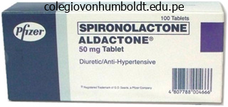
Aldactone dosages: 100 mg, 25 mg
Aldactone packs: 30 pills, 60 pills, 90 pills, 120 pills, 180 pills, 270 pills, 360 pills
Buy discount aldactone 100 mg on-line
In the absence of the ossicular system and tympanic membrane, sound waves can nonetheless travel immediately through the air of the center ear and enter the cochlea on the oval window. However, the sensitivity for listening to is then 15 to 20 decibels lower than for ossicular transmission- equivalent to a decrease from a medium to a barely perceptible voice degree. The malleus is certain to the incus by minute ligaments, so whenever the malleus moves, the incus strikes with it. The opposite finish of the incus articulates with the stem of the stapes, and the faceplate of the stapes lies in opposition to the membranous labyrinth of the cochlea in the opening of the oval window. The tip finish of the deal with of the malleus is connected to the middle of the tympanic membrane, and this level of attachment is constantly pulled by the tensor tympani muscle, which retains the tympanic membrane tensed. This rigidity permits sound vibrations on any portion of the tympanic membrane to be transmitted to the ossicles, which might not be true if the membrane have been lax. The ossicles of the middle ear are suspended by ligaments in such a method that the combined malleus and incus act as a single lever, having its fulcrum roughly at the border of the tympanic membrane. The articulation of the incus with the stapes causes the stapes to (1) push ahead on the oval window and on the cochlear fluid on the other aspect of window each time the tympanic membrane strikes inward and (2) pull backward on the fluid every time the malleus moves outward. The Auditory canal Tympanic membrane amplitude of motion of the stapes faceplate with each sound vibration is just three fourths as much because the amplitude of the handle of the malleus. The Special Senses Attenuation of Sound by Contraction of the Tensor Tympani and Stapedius Muscles. When loud sounds Basilar Spiral organ membrane of Corti Spiral ligament Vestibular membrane Scala vestibuli Stria vascularis are transmitted via the ossicular system and from there into the central nervous system, a reflex occurs after a latent interval of only 40 to 80 milliseconds to cause contraction of the stapedius muscle and, to a lesser extent, the tensor tympani muscle. The tensor tympani muscle pulls the handle of the malleus inward while the stapedius muscle pulls the stapes outward. These two forces oppose one another and thereby cause the whole ossicular system to develop elevated rigidity, thus tremendously lowering the ossicular conduction of low-frequency sound, primarily frequencies below 1000 cycles/sec. This attenuation reflex can scale back the intensity of lower-frequency sound transmission by 30 to 40 decibels, which is about the identical distinction as that between a loud voice and a whisper. The function of this mechanism is believed to be twofold: to defend the cochlea from damaging vibrations brought on by excessively loud sound and to masks low-frequency sounds in loud environments. Masking normally removes a major share of the background noise and permits an individual to think about sounds above 1000 cycles/sec, where a lot of the pertinent info in voice communication is transmitted. This impact is activated by collateral nerve indicators transmitted to these muscles at the same time that the brain prompts the voice mechanism. Therefore, under appropriate situations, a tuning fork or an electronic vibrator positioned on any bony protuberance of the cranium, but especially on the mastoid course of near the ear, causes the individual to hear the sound. It consists of three tubes coiled side by aspect: (1) the scala vestibuli, (2) the scala media, and (3) the scala tympani. On the floor of the basilar membrane lies the organ of Corti, which accommodates a series of electromechanically sensitive cells, the hair cells. They are the receptive finish organs that generate nerve impulses in response to sound vibrations. Therefore, as far as fluid conduction of sound is concerned, the scala vestibuli and scala media are thought-about to be a single chamber. Inward motion causes the fluid to move ahead within the scala vestibuli and scala media, and outward motion causes the fluid to move backward. It incorporates 20,000 to 30,000 basilar fibers that project from the bony center of the cochlea, the modiolus, toward the outer wall. The lengths of the basilar fibers enhance progressively beginning at the oval window and going from the base of the cochlea to the apex, growing from a size of about zero. The diameters of the fibers, nevertheless, lower from the oval window to the helicotrema, so their overall stiffness decreases greater than 100-fold. As a result, the stiff, short fibers near the oval window of the cochlea vibrate finest at a very excessive frequency, whereas the long, limber fibers close to the tip of the cochlea vibrate finest at a low frequency. Thus, high-frequency resonance of the basilar membrane occurs near the base, the place the sound waves enter the cochlea by way of the oval window. However, lowfrequency resonance occurs near the helicotrema, mainly due to the much less stiff fibers but additionally because of elevated "loading" with additional plenty of fluid that must vibrate along the cochlear tubules. The initial impact of a sound wave entering on the oval window is to trigger the basilar membrane at the base totally different patterns of transmission for sound waves of various frequencies.
Cheap aldactone online amex
Some sufferers with psychological despair alternate between despair and mania, which is called both bipolar dysfunction or manic-depressive psychosis, and fewer patients exhibit solely mania without the depressive episodes. Drugs that diminish the formation or action of norepinephrine and serotonin, such as lithium compounds, could be effective in treating the manic phase of the condition. In help of this idea is the reality that pleasure and reward centers of the hypothalamus and surrounding areas obtain large numbers of nerve endings from the norepinephrine and serotonin techniques. Schizophrenia-Possible Exaggerated Function of Part of the Dopamine System Schizophrenia comes in many types. One of the commonest types is seen in the particular person who hears voices and has delusions, intense concern, or other types of emotions which are unreal. Many schizophrenics are highly paranoid, with a way of persecution from exterior sources. The reason for believing that the prefrontal lobes are involved in schizophrenia is that a schizophrenic-like sample of mental activity may be induced in monkeys by making multiple minute lesions in widespread areas of the prefrontal lobes. It has been advised that in individuals with schizophrenia, extra dopamine is secreted by a group of dopaminesecreting neurons whose cell our bodies lie within the ventral tegmentum of the mesencephalon, medial and superior to the substantia nigra. These neurons give rise to the so-called mesolimbic dopaminergic system that tasks nerve fibers and dopamine secretion into the medial and anterior portions of the limbic system, particularly into the hippocampus, amygdala, anterior caudate nucleus, and portions of the prefrontal lobes. An much more compelling cause for believing that schizophrenia might be attributable to extra production of dopamine is that many medicine which might be effective in treating schizophrenia, similar to chlorpromazine, haloperidol, and thiothixene, all either decrease secretion of dopamine at dopaminergic nerve endings or lower the effect of dopamine on subsequent neurons. Finally, attainable involvement of the hippocampus in schizophrenia was found when it was realized that in individuals with schizophrenia, the hippocampus is usually shrunk, particularly in the dominant hemisphere. Motor and sensory abnormalities, gait disturbances, and seizures are uncommon until the late phases of the disease. The peptide accumulates in amyloid plaques, which vary in diameter from 10 micrometers to several hundred micrometers and are found in widespread areas of the brain, including in the cerebral cortex, hippocampus, basal ganglia, thalamus, and even the cerebellum. Cirelli C: the genetic and molecular regulation of sleep: from fruit fliestohumans. This system helps to management arterial pressure, gastrointestinal motility, gastrointestinal secretion, urinary bladder emptying, sweating, physique temperature, and heaps of other activities. Some of those actions are managed almost entirely and a few solely partially by the autonomic nervous system. One of essentially the most striking characteristics of the autonomic nervous system is the rapidity and intensity with which it could change visceral functions. For occasion, within three to 5 seconds it can increase the guts fee to twice regular, and inside 10 to 15 seconds the arterial stress could be doubled. At the opposite excessive, the arterial strain may be decreased low sufficient inside 10 to 15 seconds to trigger fainting. Sweating can begin within seconds, and the urinary bladder could empty involuntarily, also within seconds. The sympathetic nerve fibers originate in the spinal wire along with spinal nerves between twine segments T1 and L2 and move first into the sympathetic chain after which to the tissues and organs which are stimulated by the sympathetic nerves. In addition, portions of the cerebral cortex, particularly of the limbic cortex, can transmit signals to the lower facilities and on this way can influence autonomic control. That is, subconscious sensory alerts from visceral organs can enter the autonomic ganglia, the brain stem, or the hypothalamus after which return unconscious reflex responses directly back to the visceral organs to management their actions. The efferent autonomic alerts are transmitted to the various organs of the physique through two major subdivisions called the sympathetic nervous system and the parasympathetic nervous system, the characteristics and features of that are described within the following sections. Shown the sympathetic nerves are different from skeletal motor nerves in the following method: Each sympathetic pathway from the twine to the stimulated tissue consists of two neurons, a preganglionic neuron and a postganglionic neuron, in distinction to only a single neuron in the skeletal motor pathway. Immediately after the spinal nerve leaves the spinal canal, the preganglionic sympathetic fibers go away the spinal nerve and move through a white ramus into one of the ganglia of the sympathetic chain. The postganglionic sympathetic neuron thus originates either in one of the sympathetic chain ganglia or in one of many peripheral sympathetic ganglia. From either of these two sources, the postganglionic fibers then journey to their locations in the varied organs. These sympathetic fibers are all very small type C fibers, they usually lengthen to all elements of the physique by the use of the skeletal nerves. Nerve connections among the many spinal wire, spinal nerves, sympathetic chain, and peripheral sympathetic nerves.
Purchase aldactone 25 mg visa
For instance, subconscious control of arterial stress and respiration is achieved primarily within the medulla and pons. Control of equilibrium is a mixed perform of the older portions of the cerebellum and the reticular substance of the medulla, pons, and mesencephalon. In addition, many emotional patterns similar to anger, pleasure, sexual response, response to pain, and response to pleasure can still occur after destruction of a lot of the cerebral cortex. The answer to this query is complex, but it begins with the fact that the cerebral cortex is a particularly large memory storehouse. The cortex never features alone but always in association with decrease centers of the nervous system. Without the cerebral cortex, the capabilities of the lower mind facilities are often imprecise. The huge storehouse of cortical info often converts these capabilities to determinative and precise operations. However, as well as, every impulse (1) could additionally be blocked in its transmission from one neuron to the subsequent, (2) could additionally be changed from a single impulse into repetitive impulses, or (3) may be integrated with impulses from different neurons to cause extremely intricate patterns of impulses in successive neurons. First, all computer systems have enter circuits that may be compared with the sensory portion of the nervous system, as properly as output circuits which are analogous to the motor portion of the nervous system. In simple computers, the output indicators are managed immediately by the input alerts, working in a manner much like that of easy reflexes of the spinal wire. In more complex computers, the output is decided both by enter alerts and by data that has already been stored in reminiscence in the pc, which is analogous to the extra complicated reflex and processing mechanisms of our greater nervous system. This unit is analogous to the control mechanisms in our mind that direct our attention first to one thought or sensation or motor exercise, then to another, and so forth, until complex sequences of thought or motion take place. Even a rapid study of this diagram demonstrates its simi larity to the nervous system. Most of the synapses used for signal transmission in the central nervous system of the human being are chemical synapses. In these synapses, the first neuron secretes at its nerve ending synapse a chemical substance referred to as a neurotransmitter (often called a transmitter substance), and this transmitter in flip acts on receptor proteins within the membrane of the next neuron to excite the neuron, inhibit it, or modify its sensitivity in another way. In electrical synapses, the cytoplasms of adjacent cells are immediately related by clusters of ion channels referred to as hole junctions that allow free movement of ions from the inside of one cell to the inside of the next cell. Chem Presynaptic terminal Neurotransmitter Synaptic cleft (200-300 �) Ionotropic receptor Postsynaptic terminal Metabotropic receptor Ions Second messenger Cellular response: � Membrane potential � Biochemical cascades � Regulation of gene expression B Electrical synapse Action potential ical synapses have one exceedingly necessary character istic that makes them highly desirable for transmitting nervous system alerts. This attribute is that they at all times transmit the alerts in a single direction-that is, from the neuron that secretes the neurotransmitter, referred to as the presynaptic neuron, to the neuron on which the transmit ter acts, called the postsynaptic neuron. A oneway conduction mechanism permits alerts to be directed towards particular goals. It is composed of three major components: the soma, which is the primary body of the neuron; a single axon, which extends from the soma right into a peripheral nerve that leaves the spinal twine; and the dendrites, that are great numbers of branching projec tions of the soma that stretch as a lot as 1 millimeter into the encompassing areas of the wire. As many as 10,000 to 200,000 minute synaptic knobs referred to as presynaptic terminals lie on the surfaces of the dendrites and soma of the motor neuron, with about eighty to 95 % of them on the dendrites and only 5 to 20 % on the soma. These presynaptic terminals are the ends of nerve fibrils that originate from many different neurons. Many of those presynaptic terminals are excitatory-that is, they secrete a neurotransmitter that excites the postsynaptic neuron. However, different presyn aptic terminals are inhibitory-that is, they secrete a neu rotransmitter that inhibits the postsynaptic neuron. Neurons in different components of the wire and mind differ from the anterior motor neuron in (1) the dimensions of the cell physique; (2) the size, dimension, and variety of dendrites, ranging in size from almost zero to many centimeters; (3) the size and size of the axon; and (4) the number of presynaptic terminals, which can range from only some to as many as 200,000. These variations make neurons in several components of the nervous system react in one other way to incoming synaptic alerts and, due to this fact, perform many alternative capabilities. Although most synapses within the brain are chemical, electrical and chemical synapses might coexist and interact within the central nervous system. The bidirectional transmis sion of electrical synapses permits them to help coor dinate the activities of large groups of interconnected neurons. General Principles and Sensory Physiology membrane, which ends up in excitation or inhibition of the postsynaptic neuron, relying on the neuronal receptor traits. The presyn aptic terminal is separated from the postsynaptic neuro nal soma by a synaptic cleft having a width often of 200 to 300 angstroms. The terminal has two internal struc tures essential to the excitatory or inhibitory operate of the synapse: the transmitter vesicles and the mitochondria. The transmitter vesicles comprise the neurotransmitter that, when released into the synaptic cleft, either excites or inhibits the postsynaptic neuron.
Purchase 100 mg aldactone overnight delivery
On each side of the brain, these ganglia consist of the caudate nucleus, putamen, globus pallidus, substantia nigra, and subthalamic nucleus. Anatomical relations of the basal ganglia to the cerebral cortex and thalamus, shown in three-dimensional view. Putamen circuit through the basal ganglia for unconscious execution of learned patterns of movement. When the basal ganglia sustain severe damage, the cortical system of motor management can no longer present these pat terns. Other patterns that require the basal ganglia are cutting paper with scissors, hammering nails, taking pictures a basket ball via a hoop, passing a football, throwing a base ball, the movements of shoveling dust, most features of vocalization, controlled movements of the eyes, and vir tually any other of our skilled movements, most of them performed subconsciously. Relation of the basal ganglial circuitry to the corticospinal-cerebellar system for motion control. Almost all motor and sensory nerve fibers connecting the cere bral cortex and spinal wire cross through the house that lies between the most important plenty of the basal ganglia, the caudate nucleus and the putamen. It is important for our present dialogue due to the intimate affiliation between the basal ganglia and the corticospinal system for motor control. To the left is proven the motor cortex, thalamus, and associated brain stem and cerebellar circuitry. To the best is the major circuitry of the basal ganglia system, displaying the tremendous interconnections among the basal ganglia plus intensive enter and output pathways between the other motor regions of the mind and the basal ganglia. In the subsequent few sections we focus particularly on two main circuits, the putamen circuit and the caudate circuit. They start mainly within the premotor and supplementary areas of the motor cortex and in the somatosensory areas of the sensory cortex. Next they pass to the putamen (mainly bypassing the caudate nucleus), then to the inner portion of the globus pallidus, and next to the ventroan terior and ventrolateral relay nuclei of the thalamus, and they lastly return to the cerebral primary motor cortex and to parts of the premotor and supplementary cere bral areas closely associated with the primary motor cortex. Thus, the putamen circuit has its inputs mainly from the components of the brain adjacent to the primary motor cortex but not a lot from the primary motor cortex itself. Motor and Integrative Neurophysiology cortex or closely related premotor and supplementary cortex. Functioning in close affiliation with this primary putamen circuit are ancillary circuits that move from the putamen via the exterior globus pallidus, the subthalamus, and the substantia nigra-finally returning to the motor cortex by means of the thalamus. How does the putamen Premotor and supplemental Prefrontal Primary motor Somatosensory circuit operate to help execute patterns of motion However, when a portion of the circuit is damaged or blocked, sure patterns of motion turn out to be severely irregular. For instance, lesions in the globus pallidus frequently lead to spontaneous and sometimes continuous writhing actions of a hand, an arm, the neck, or the face. A lesion in the subthalamus often results in sudden flailing movements of a complete limb, a situation referred to as hemiballismus. Multiple small lesions in the putamen result in flicking movements within the arms, face, and different components of the body, referred to as chorea. Caudate circuit through the basal ganglia for cognitive planning of sequential and parallel motor patterns to achieve specific conscious objectives. Most of our motor actions occur as a consequence of thoughts generated in the mind, a course of called cognitive management of motor exercise. The caudate nucleus plays a significant role on this cognitive control of motor activity. Furthermore, the caudate nucleus receives large amounts of its enter from the affiliation areas of the cerebral cortex overlying the caudate nucleus, primarily areas that additionally integrate the several sorts of sensory and motor data into usable thought patterns. Instead, the returning alerts go to the accessory motor regions in the premotor and supple mentary motor areas which may be concerned with placing collectively sequential patterns of motion lasting 5 or more seconds as an alternative of exciting individual muscle actions. A good instance of this phenomenon can be a person seeing a lion method after which responding instantaneously and routinely by (1) turning away from the lion, (2) beginning to run, and (3) even try ing to climb a tree. Thus, cognitive management of motor exercise determines subconsciously, and within seconds, which patterns of movement will be used collectively to achieve a posh objective that might itself final for a lot of seconds. Also, he or she may write a small "a" on a chunk of paper or a big "A" on a chalkboard. Regardless of the selection, the proportional traits of the letter remain almost the same.
Purchase aldactone without prescription
The presence of hemoglobin within the red blood cells allows the blood to transport 30 to one hundred instances as much O2 as might be transported within the form of dissolved O2 within the water of the blood. Therefore, the preliminary strain difference that causes O2 to diffuse into the pulmonary capillary is 104 - forty, or 64 mm Hg. Also, due to elevated cardiac output during train, the time that the blood stays in the pulmonary capillary may be lowered to lower than onehalf regular. Yet because of the good safety factor for diffusion of O2 through the pulmonary membrane, the blood nonetheless becomes almost saturated with O2 by the point it leaves the pulmonary capillaries. First, it was pointed out in Chapter 40 that the diffusing capacity for O2 will increase nearly threefold throughout exercise; this results mainly from elevated floor area of capillaries collaborating in the diffusion and also from a extra nearly best ventilation-perfusion ratio within the higher part of the lungs. That is, the blood normally stays within the lung capillaries about 3 times so lengthy as wanted to cause full oxygenation. Therefore, throughout exercise, even with a shortened time of publicity in the capillaries, the blood can still turn into virtually absolutely oxygenated. This blood circulate is called "shunt flow," which means that blood is shunted previous the gasoline change areas. In the lungs, it diffuses from the pulmonary capillaries into the alveoli and is expired. The remaining three % is transported in the dissolved state in the water of the plasma and blood cells. Thus, under normal conditions, oxygen is carried to the tissues nearly completely by hemoglobin. Amount of Oxygen Released From the Hemoglobin When Systemic Arterial Blood Flows Through the Tissues. The complete quantity of O2 certain with hemoglobin in regular systemic arterial blood, which is ninety seven p.c saturated, is about 19. Upon passing by way of the tissue capillaries, this amount is reduced, on average, to 14. Thus, under normal conditions, about 5 milliliters of O2 are transported from the lungs to the tissues by each one hundred milliliters of blood move. Keep in mind that the cardiac output can increase to six to seven occasions regular in well-trained marathon runners. Thus, multiplying the rise in cardiac output (6- to 7-fold) by the increase in O2 transport in every volume of blood (3-fold) offers a 20-fold enhance in O2 transport to the tissues. The percentage of the blood that gives up its O2 as it passes by way of the tissue capillaries is recognized as the utilization coefficient. The regular worth for that is about 25 percent, as is obvious from the previous discussion-that is, 25 p.c of the oxygenated hemoglobin offers its O2 to the tissues. During strenuous exercise, the utilization coefficient in the entire body can improve to 75 to 85 %. In native tissue areas the place blood flow is extremely sluggish or the metabolic fee may be very high, utilization coefficients approaching 100% have been recorded-that is, essentially all of the O2 is given to the tissues. Conversely, during heavy train, extra amounts of O2 (as a lot as 20 times normal) should be delivered from the hemoglobin to the tissues. Only a small amount of additional O2 dissolves in the fluid of the blood, as shall be mentioned subsequently. This determine shows that when the blood becomes barely acidic, with the pH decreasing from the conventional value of seven. Shift of the oxygen-hemoglobin dissociation curve to the best brought on by an increase in hydrogen ion focus (decreaseinpH). All these factors act together to shift the oxygen-hemoglobin dissociation curve of the muscle capillary blood significantly to the best. Then, in the lungs, the shift occurs in the reverse direction, permitting the pickup of extra amounts of O2 from the alveoli. Only a minute level of O2 strain is required in the cells for regular intracellular chemical reactions to happen. If the rate of blood flow falls to zero, the quantity of obtainable O2 also falls to zero. Thus, there are occasions when the speed of blood flow through a tissue could be so low that tissue Po2 falls under the important 1 mm Hg required for intracellular metabolism. Neither diffusion-limited nor blood flow�limited oxygen states can proceed for long, as a outcome of the cells obtain much less O2 than is required to continue the life of the cells. This figure compares with virtually 5 milliliters of O2 transported by the purple blood cell hemoglobin.
Purchase aldactone on line
Fibers from most discrete cutaneous tactile receptors and from the flowerspray endings of the muscle spindles (about eight micrometers in diameter on common; these fibers are and kind A fibers in the general classification). Unmyelinated fibers carrying pain, itch, temperature, and crude touch sensations (0. The different gradations of intensity may be trans mitted both by utilizing growing numbers of parallel fibers or by sending more motion potentials alongside a single fiber. These two mechanisms are known as, respectively, spatial summation and temporal summation. This determine exhibits a piece of pores and skin innervated by numerous parallel pain fibers. Each of those fibers arborizes into hundreds of minute free nerve endings that function pain receptors. The entire cluster of fibers from one pain fiber regularly covers an area of skin as massive as 5 centimeters in diameter. The number of endings is massive in the middle of the sphere but diminishes toward the periphery. One also can see from the figure that the arborizing fibrils overlap these from different ache fibers. Therefore, a pinprick of the pores and skin often stimulates endings from many various pain fibers simultaneously. To the left is the impact of a weak stimulus, with only a single nerve fiber in the midst of the bundle stimulated strongly (represented by the redcolored fiber), whereas a quantity of adjoining fibers are stimulated weakly (halfred fibers). The different two views of the nerve cross part present the effect of a reasonable stimulus and a robust stimulus, with progressively more fibers being stimulated. A second means for transmit ting signals of accelerating power is by growing the frequency of nerve impulses in every fiber, which is recognized as temporal summation. For occasion, the complete cerebral cortex might be considered to be a single large neuronal pool. Other neuronal swimming pools embrace the different basal ganglia and the specific nuclei within the thalamus, cerebellum, mesencephalon, pons, and medulla. Also, the whole dorsal gray matter of the spinal twine could presumably be considered one long pool of neurons. Each neuronal pool has its own particular organization that causes it to process alerts in its own distinctive method, thus allowing the entire consortium of swimming pools to obtain the multitude of functions of the nervous system. Yet, despite their differences in perform, the swimming pools also have many comparable rules of function, described in the following sections. The neuronal space stimulated by each incoming nerve fiber is identified as its stimulatory subject. Note that enormous numbers of the terminals from each input fiber lie on the closest neuron in its "area," however progressively fewer terminals lie on the neurons farther away. Instead, giant numbers of input terminals should discharge on the same neuron either concurrently or in fast succession to trigger excitation. Note that input fiber 1 has greater than sufficient terminals to trigger neuron a to discharge. Input fiber 1 additionally contributes terminals to neurons b and c, however not enough to trigger excitation. Nevertheless, discharge of these terminals makes both these neurons extra likely to be excited by signals arriving through other incoming nerve fibers. Therefore, the stimuli to these neurons are mentioned to be subthreshold, and the neurons are stated to be facilitated. Similarly, for enter fiber 2, the stimulus to neuron d is a suprathreshold stimulus, and the stimuli to neurons b and c are subthreshold, but facilitating, stimuli. In the central portion of the sector in this figure, designated by the circled area, all the neurons are stimulated by the incoming fiber. Therefore, this is said to be the discharge zone of the incoming fiber, additionally known as the excited zone or liminal zone.
Cheap aldactone 25 mg amex
Thromboplastin, which is critical to provoke the clotting course of, consists primarily of one of many cephalins. Large portions of sphingomyelin are current in the nervous system; this substance acts as an electrical insulator within the myelin sheath round nerve fibers. Phospholipids are donors of phosphate radicals when these radicals are necessary for different chemical reactions within the tissues. Perhaps the most important of all the capabilities of phospholipids is participation within the formation of structural elements-mainly membranes-in cells throughout the body, as mentioned in the subsequent section of this chapter in connection with an analogous function for ldl cholesterol. Essentially all of the endogenous ldl cholesterol that circulates within the lipoproteins of the plasma is fashioned by the liver, but all other cells of the body kind no less than some ldl cholesterol, which is according to the reality that lots of the membranous structures of all cells are partially composed of this substance. The basic construction of ldl cholesterol is a sterol nucleus, which is synthesized completely from multiple molecules of acetyl-CoA. In turn, the sterol nucleus could be modified by various aspect chains to form (1) ldl cholesterol; (2) cholic acid, which is the idea of the bile acids formed in the liver; and (3) many essential steroid hormones secreted by the adrenal cortex, the ovaries, and the testes (these hormones are discussed in later chapters). Factors That Affect Plasma Cholesterol Concentration- Feedback Control of Body Cholesterol. Indeed, about 70 percent of the ldl cholesterol within the lipoproteins of the plasma is within the type of cholesterol esters. An enhance within the quantity of ldl cholesterol ingested every day could enhance the plasma concentration barely. However, when ldl cholesterol is ingested, the rising focus of cholesterol inhibits probably the most essential enzyme for endogenous synthesis of cholesterol, 3-hydroxy-3-methylglutaryl CoA reductase, thus providing an intrinsic suggestions control system to prevent an extreme improve in plasma ldl cholesterol concentration. A food plan excessive in saturated fats increases blood cholesterol concentration 15 to 25 percent, particularly when this food regimen is associated with excess weight gain and obesity. This improve in blood ldl cholesterol results from elevated fat deposition in the liver, which then offers elevated portions of acetyl-CoA in the liver cells for the manufacturing of cholesterol. Therefore, to lower the blood cholesterol concentration, sustaining a diet low in saturated fat and a normal physique weight is much more necessary than sustaining a food regimen low in cholesterol. Ingestion of fats containing highly unsaturated fatty acids often depresses the blood ldl cholesterol concentration a slight to reasonable quantity. The mechanism of this impact is unknown, although this remark is the idea of much present-day dietary strategy. Lack of insulin or thyroid hormone will increase the blood cholesterol concentration, whereas extra thyroid hormone decreases the focus. These effects are in all probability caused primarily by changes in the degree of activation of specific enzymes answerable for the metabolism of lipid substances. Genetic problems of ldl cholesterol metabolism might significantly improve plasma levels of cholesterol. As mentioned later, this phenomenon causes the liver to produce extreme amounts of cholesterol. By far essentially the most abundant non-membranous use of ldl cholesterol within the body is to kind cholic acid within the liver. As explained in Chapter 71, cholic acid is conjugated with different substances to form bile salts, which promote digestion and absorption of fat. A small amount of ldl cholesterol is used by (1) the adrenal glands to kind adrenocortical hormones, (2) the ovaries to form progesterone and estrogen, and (3) the testes to kind testosterone. These glands can also synthesize their own sterols after which kind hormones from them, as mentioned in the chapters on endocrinology. This cholesterol, along with other lipids, makes the skin extremely immune to the absorption of water-soluble substances and to the motion of many chemical brokers because ldl cholesterol and the other skin lipids are extremely inert to acids and to many solvents which may in any other case easily penetrate the body. Cellular Structural Functions of Phospholipids and Cholesterol-Especially for Membranes the previously talked about uses of phospholipids and ldl cholesterol are of only minor importance in comparison with their perform of forming specialized constructions, mainly membranes, in all cells of the body. In Chapter 2, it was pointed out that giant portions of phospholipids and ldl cholesterol are present in each the cell membrane and the membranes of the interior organelles of all cells. It can be identified that the ratio of membrane ldl cholesterol to phospholipids is very essential in figuring out the fluidity of cell membranes. Thus, the bodily integrity of cells all over the place within the physique relies mainly on phospholipids, cholesterol, and certain insoluble proteins. The polar charges on the phospholipids also reduce the interfacial rigidity between the cell membranes and the encompassing fluids.
Aldactone 25 mg purchase without a prescription
Unlike the cortical amassing tubule, the medullary amassing duct is permeable to urea, and there are particular urea transporters that facilitate urea diffusion across the luminal and basolateral membranes. The medullary accumulating duct is capable of secreting hydrogen ions in opposition to a large focus gradient, as also happens within the cortical accumulating tubule. Thus, the medullary accumulating duct additionally plays a key position in regulating acid-base stability. If a larger proportion of water is reabsorbed, the substance turns into extra concentrated. If a larger proportion of the solute is reabsorbed, the substance turns into more diluted. All the values on this determine represent the tubular fluid concentration divided by the plasma concentration of a substance. If plasma focus of the substance is assumed to be constant, any change in the tubular fluid/ plasma focus ratio reflects changes in tubular fluid concentration. As the filtrate moves along the tubular system, the concentration rises to progressively greater than 1. An necessary characteristic of tubular reabsorption is that reabsorption of some solutes may be regulated independently of others, particularly by way of hormonal control mechanisms. Conversely, the substances represented toward the bottom of the figure, corresponding to glucose and amino acids, are all strongly reabsorbed; these are all substances that the body must conserve, and nearly none of them are lost within the urine. Tubular Fluid/Plasma Inulin Concentration Ratio Can Be Used to Measure Water Reabsorption by the Renal Tubules. Changes in inulin concentration at totally different points alongside the renal tubule, therefore, mirror adjustments in the amount of water present in the tubular fluid. For example, the tubular fluid/plasma focus ratio for inulin rises to about three. Some diploma of glomerulotubular balance additionally happens in different tubular segments, especially the loop of Henle. It is evident that the mechanisms for glomerulotubular steadiness can happen independently of hormones and could be demonstrated in completely isolated kidneys and even in fully isolated proximal tubular segments. Changes in peritubular capillary reabsorption can in flip affect the hydrostatic and colloid osmotic pressures of the renal interstitium and, in the end, reabsorption of water and solutes from the renal tubules. Fluid and electrolytes are reabsorbed from the tubules into the renal interstitium and from there into the peritubular capillaries. Reabsorption throughout the peritubular capillaries may be calculated as Reabsorption = K f � Net reabsorptive drive the web reabsorptive drive represents the sum of the hydrostatic and colloid osmotic forces that both favor or oppose reabsorption across the peritubular capillaries. These forces include (1) hydrostatic strain inside the peritubular capillaries (peritubular hydrostatic stress [Pc]), which opposes reabsorption; (2) hydrostatic pressure in the renal interstitium (Pif) exterior the capillaries, which favors reabsorption; (3) colloid osmotic stress of the peritubular capillary plasma proteins (c), which favors reabsorption; and (4) colloid osmotic strain of the proteins within the renal interstitium (if), which opposes reabsorption. This opposition to fluid reabsorption is greater than counterbalanced by the colloid osmotic pressures that favor reabsorption. The plasma colloid osmotic 360 stress, which favors reabsorption, is about 32 mm Hg, and the colloid osmotic stress of the interstitium, which opposes reabsorption, is 15 mm Hg, causing a net colloid osmotic drive of about 17 mm Hg, favoring reabsorption. Therefore, subtracting the net hydrostatic forces that oppose reabsorption (7 mm Hg) from the web colloid osmotic forces that favor reabsorption (17 mm Hg) gives a net reabsorptive pressure of about 10 mm Hg. This worth is excessive, just like that discovered in the glomerular capillaries, however in the incorrect way. The different factor that contributes to the high price of fluid reabsorption within the peritubular capillaries is a big filtration coefficient (Kf) due to the excessive hydraulic conductivity and large surface space of the capillaries. Because the reabsorption fee is normally about 124 ml/min and net reabsorption pressure is 10 mm Hg, Kf normally is about 12. The two determinants of peritubular capillary reabsorption which would possibly be instantly influenced by renal hemodynamic adjustments are the hydrostatic and colloid osmotic pressures of the peritubular capillaries. The peritubular capillary hydrostatic pressure is influenced by the arterial pressure and resistances of the afferent and efferent arterioles as follows. This effect is buffered to some extent by autoregulatory mechanisms that keep relatively fixed renal blood flow, as well as comparatively constant hydrostatic pressures in the renal blood vessels. The second major determinant of peritubular capillary reabsorption is the colloid osmotic pressure of the plasma in these capillaries; elevating the colloid osmotic strain will increase peritubular capillary reabsorption. The colloid osmotic strain of peritubular capillaries is determined by (1) the systemic plasma colloid osmotic pressure (increasing the plasma protein concentration of systemic blood tends to raise peritubular capillary colloid osmotic strain, thereby growing reabsorption) and (2) the filtration fraction (the greater the filtration fraction, the greater the fraction of plasma filtered by way of the glomerulus and, consequently, the extra concentrated the protein becomes in the plasma that remains behind). Thus, rising the filtration fraction also tends to enhance the peritubular capillary reabsorption fee.
Real Experiences: Customer Reviews on Aldactone
Yorik, 44 years: The partial pressure represents a measure of the total number of molecules of a particular fuel hanging a unit space of the alveolar surface of the membrane in unit time, and the stress of the gas within the blood represents the variety of molecules that try and escape from the blood in the incorrect way.
Frithjof, 63 years: The forms of components which are frequently monitored in the duodenum and might provoke enterogastric inhibitory reflexes embody the following: 1.
Bernado, 24 years: The hepatic cells include giant quantities of a protein known as apoferritin, which is able to combining reversibly with iron.
Pakwan, 58 years: If a diver has been beneath the ocean long enough that enormous amounts of nitrogen have dissolved in his or her body and the diver then all of a sudden comes again to the floor of the sea, significant quantities of nitrogen bubbles can develop in the body fluids either intracellularly or extracellularly and might cause minor or severe injury in nearly any area of the physique, relying on the quantity and sizes of bubbles formed; this phenomenon known as decompression sickness.
Silvio, 45 years: Vitamin E is believed to play a protecting function within the prevention of oxidation of unsaturated fats.
Ortega, 50 years: Thus, easy command signals from the brain can initiate many normal motor activities, particu larly for such features as strolling and attaining totally different postural attitudes of the physique.
Marik, 27 years: General Principles and Sensory Physiology being recognized in 10 to 20 gradations of strength, quite than as many as a hundred gradations for the dorsal column system; and (4) the power to transmit rapidly altering or quickly repetitive signals is poor.
Kaffu, 42 years: Maintenance of steadiness between intake and output of potassium depends totally on excretion by the kidneys because the quantity excreted within the feces is just about 5 to 10 % of the potassium consumption.
10 of 10 - Review by G. Connor
Votes: 42 votes
Total customer reviews: 42
References
- Slim K, Vicaut E, Launay-Savary M-V, et al: Updated systematic review and meta-analysis of randomized clinical trials on the role of mechanical bowel preparation before colorectal surgery, Ann Surg 249:203n209, 2009.
- Breithardt OA, Stellbrink C, Herbots L, et al: Cardiac resynchronization therapy can reverse abnormal myocardial strain distribution in patients with heart failure and left bundle branch block, J Am Coll Cardiol 42:486-494, 2003.
- Chrysos E, Athanasakis E, Pechlivanides G, et al: The effect of total and anterior partial fundoplication on antireflux mechanisms of the gastroesophageal junction. Am J Surg 188:39, 2004.
- Lloyd EA, Gersh BJ, Kennelly BM. Hemodynamic spectrum of ' 'dominant' right ventricular infarction in 19 patients. Am J Cardiol. 1981;48(6):1016-1022.
- Agrawal, M.S., Agrawal, M., Gupta, A., Bansal, S., Yadav, A., Goyal, J. A randomized comparison of tubeless and standard percutaneous nephrolithotomy. J Endourol 2008;22:439-442.
- Ordonez NG. Utilization of thyroid transcription factor-1 immunostaining in the diagnosis of lung tumors. Methods Mol Med 2003;75:355-68.


