


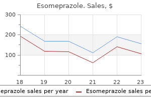
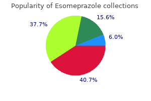
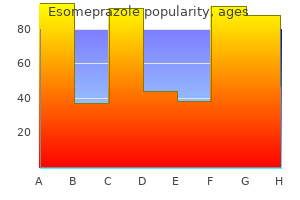
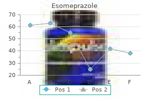
Esomeprazole
Esomeprazole dosages: 40 mg, 20 mg
Esomeprazole packs: 60 pills, 90 pills, 120 pills, 180 pills, 270 pills, 360 pills

40 mg esomeprazole with visa
Such a pattern indicates delayed drainage without parenchymal dysfunction and is nearly at all times with none scientific significance. The possibility of a vesicoureteric reflux of tracer through the drainage part also needs to be saved in mind so as to be in a position to differentiate it from outflow tract obstruction. The background exercise is often a mirrored image of blood clearance of tracer or quite the renal extraction of tracer from the blood pool, which quickly decreases with time in a affected person with normal renal operate. An extra picture is recommended to determine the tracer clearance, which, if enough, should be considered normal, and the outflow tract not obstructed. Normal Dynamic Renal Study There is an intense and rapid focus of tracer in the renal parenchyma at 1�3 minutes postinjection. Ureters may be visualized in normal patients and in these with sluggish ureteral transit time. The renal Diuretic Renography Furosemide causes a fast diuretic response that drains out tracer from dilated nonobstructed system. In significant outflow tract obstruction, tracer within the renal space could decrease slowly or fail to lower or even enhance in response to a diuretic problem. Normal time exercise curves with steep uptake slope, distinct peak, and rapid clearance with normal early and delayed static photographs (C) Chapter 97 Current Status of Nuclear Medicine in Urinary Tract Imaging 1555 but also on the power of the parenchyma to reply to a diuretic. Drainage is directly proportional to urine flow and inversely proportional to the amount of the renal pelvis and ureters. Compliance and quantity of the pelvicalyceal system are also essential in that a large flaccid renal pelvis usually causes slow drainage of radiotracer. Severe hydroureteronephrosis could partly or wholly masks the impact of a diuretic on the emptying of the renal pelvis. Other elements embody poor response of the renal parenchyma to the diuretic, poor supply of the diuretic to the kidneys, bladder over distension, complicated pelvic surgical procedures, prune-belly syndrome and ectopic kidney. Imaging: Diuretic renography could be carried out utilizing the identical radiopharmaceuticals talked about earlier. It is crucial to be sure that tracer has filled the renal pelvis and postural drainage has not occurred earlier than the diuretic is run. The diuretic could be administered quarter-hour postinjection or concurrently with the radiotracer. Forced hydration is recommended as a way to differentiate obstruction from nonobstruction in diuretic renography. The diuretic impact is usually seen within 1�2 minutes after administration of furosemide. Patients with intermittent hydronephrosis who complain of intermittent flank ache could have copy of ache on administration of diuretic. In some circumstances with otherwise balanced drainage and urinary flow, a diuretic could cause speedy over distension of the renal pelvis resulting within the disruption of the status of the system. It is important to observe that in patients with urinary diversions, even within the absence of obstruction, circulate dynamics may be sluggish, making the prognosis of obstruction troublesome. Overlap of kidney and an ileal or colonic diversion may give rise to confusing images. Imaging in varied projections, elimination or substitute of exterior urine accumulating devices and makes an attempt at postural drainage might generally be useful. Interpretation: Careful evaluation of the dynamic renal examine previous a diuretic administration is necessary throughout interpretation. Usually, the pictures obtained previous to and after a diuretic injection are compared. The diuretic washout T 1/2 is estimated by utilizing an exponential interpolation between the point on the initial descent of the time activity curve and another point on the down slope while the curve is decaying monotonically. The diuretic T1/2 ought to be only one of the a quantity of elements, that are thought-about whereas assessing urinary obstruction. Although there may be many variations within the time of diuretic injection and subsequent time of acquisition following a diuretic response, the general sensitivity of a diuretic renography for the detection of obstruction in youngsters has been estimated at 93%.
Purchase esomeprazole with mastercard
Common sites of muscle contusion concerning the hip joint embrace the gluteal region and proximal thigh. Muscle Strain-Myotendinous Strain Strain accidents have an result on the myotendinous junction, the weakest level of the musculotendinous unit. The commonest strained muscle tissue about the hip joint are the rectus femoris and the hamstrings. Magnetic resonance imaging is used for preoperative assessment of the extent of retraction. The pelvic ring and proximal femur are the sites of origin or insertion for a number of major muscle groups, including the abdominal, gluteal, adductors, hamstrings and iliopsoas muscles. The injuries happen because of blunt direct trauma or secondary to oblique trauma from excessive tension or forceful contraction. The injuries can be categorized on the premise of the mechanism of trauma and the positioning of involvement. These include tendinous avulsion damage, muscle contusion and myotendinous strain. Gluteus Medius Tendon Tear-Greater Trochanteric Pain Syndrome the rotator cuff lesions of the hip contain tears of the abductor musculature, specifically the gluteus medius and minimus. It extra commonly impacts middle-aged to aged women who complain of persistent pain around hip or groin region, mimicking intra-articular hip pathology. This increased sign is taken into account to be the most particular and correct for a tear. The increased signal lateral to the higher trochanter more than likely represents sub gluteus maximus bursitis. Labral tear is now being recognized as a initial point for the beginning of degenerative joint illness. The torn labral fragment will get separated from the acetabular rim and loses its capability for cushioning and protecting the adjoining articular cartilage. Repetitive impaction by the femoral head on the acetabulum leads to the development of chondral defects and progressive arthritis. Acetabular labral tears are seen within the setting of osteoarthritis and congenital hip dysplasia, continual stress from athletic occasions or a traumatic event could lead to presence of tear. Tears of the labrum are identified each time distinction material extends into the labral substance or undermines its base at the interface between fibrocartilage Chapter 195 Magnetic Resonance Imaging of Hip and Pelvis 3229 and articular cartilage. There may be deformity within the contour of the labrum (loss of regular triangular shape). A labral tear might result in the formation of a paralabral cyst when the tear passes through the capsule allowing the joint fluid to leak. Paralabral cysts are more medial in location to the iliopsoas bursae and contiguous with the acetabular rim. In setting of osteoarthritis and congenital hip dysplasia, the superior acetabular labrum is most incessantly degenerated and torn, whereas in posterior dislocation complicated labral tears may be seen. This relationship is essential to perceive as the traditional excessive signal between the two low sign intensity buildings might mimic a labral tear. The remedy of acetabular labral tears is conservative within the initial 4�6 weeks with restricted weight bearing and in some circumstances by steroid injection. The sciatic nerve can break up via piriformis, and a portion of the nerve may be superficial to the piriformis muscle. Due to this variation in location of the sciatic nerve, compression, hypertrophy or harm to the piriformis muscle can cause irritation to the sciatic nerve, mimicking radicular signs of disk disease. Magnetic resonance imaging may show asymmetry in dimension of 1 piriformis in comparability with reverse facet. The iliopsoas bursa is the largest bursa in the physique and communicates with the hip joint in 15% of inhabitants. The iliopsoas bursa is situated anterior to the hip joint and adjoining to femoral vessels. Asymptomatic bursal fluid might happen in the presence of joint effusion as a outcome of communication with the joint. Greater trochanteric bursitis is another reason for hip ache, with patient localizing the pain to lateral facet of the hip; this results from repetitive hip flexion. Clinically, it is in all probability not attainable to differentiate bursitis from gluteus medius/ minimus tears.
Order esomeprazole 40 mg online
Their attribute location, geographic/wedge shape configuration with angulated margins, lack of mass impact, presence of normal blood vessels coursing by way of them and enhancement just like regular liver help differentiate these from true lesions. Severe continual hepatitis causes coarsening of parenchymal echotexture and increased echogenicity resulting in poor visualization of portal venous branches. Hepatomegaly, gallbladder wall thickening, and periportal hypodensity could be seen in acute hepatitis and periportal lymphadenopathy is often seen in chronic energetic hepatitis. Likewise, heterogeneous contrast enhancement and T2 hyperintensity signify extreme ongoing irritation in continual hepatitis, while absence of patchy enhancement suggests low inflammatory response. Unlike acute viral hepatitis, liver echogenicity is elevated in acute alcoholic hepatitis as a result of presence of concomitant fatty infiltration. Imaging is comparatively nonspecific but can help to exclude biliary obstruction in acute hepatitis. Clinically, onset is 2�6 weeks after remedy and is usually seen in patients receiving dose of more than 35 Gy to the liver. Severe acute hepatitis may end in decreased parenchymal echogenicity in opposition to which the portal vein branches appear brighter than regular. Because such iron is deposited primarily in the reticuloendothelial system (liver, spleen, lymph nodes, bone marrow) organ operate is usually preserved. Hemochromatosis then again, is a extra extreme form of iron accumulation (body stores up to 50-60 g iron) that adversely affects organ operate. Primary hemochromatosis is an autosomal recessive metabolic disorder of extreme iron absorption in duodenum/jejunum. The causes of secondary hemochromatosis embrace excess iron intake, anemia because of ineffective erythropoiesis requiring multiple blood 1332 Section three Gastrointestinal and Hepatobiliary Imaging transfusions, congenital transferrin deficiency, portocaval shunts and alcoholic cirrhosis. The excess iron in hemochromatosis is stored as ferritin and hemosiderin in varied organs including liver, pancreas, myocardium, joints, endocrine glands and pores and skin, in decreasing order of severity. The liver is the first organ to be broken in hemochromatosis leading to hepatomegaly in 95% of symptomatic sufferers. Fibrosis is maximal in periportal areas resulting in micronodular pigment cirrhosis. Iron induced interstitial fibrosis in the pancreas affects endocrine features to a higher extent than exocrine capabilities. Skin hyperpigmentation and diabetes mellitus results in so known as bronze diabetes in these patients. Splenomegaly is current in approximately 50% cases, arthropathy of small joints of the hand with or without proof of chondrocalcinosis on radiographs happens in 20�50% and cardiac involvement is the presenting criticism in 15% circumstances. Diminished sexual exercise, lack of body hair and testicular atrophy may be seen as a end result of decreased production of gonadotropins. The disease responds favorably to remedy that features venesection/removal of blood and iron chelators. Ultrasonography Ultrasonography solely depicts nonspecific changes of cirrhosis in patients with hemochromatosis and due to this fact, has no position in the diagnosis. The increased background liver attenuation in hemochromatosis might hamper detection of hepatocellular carcinoma within the postcontrast pictures but facilitates tumor detection in unenhanced scans. Magnetic Resonance Imaging Magnetic resonance imaging is probably the most delicate and specific imaging modality for demonstration of hepatic iron overload and likewise for follow-up of patients underneath treatment. Decreased sign depth in hemochromatosis can be demonstrated in pancreas and myocardium, and only sometimes in spleen. It might happen with out preexisting disease or may be seen as a complication of multiple myeloma. Normal signal depth of the pancreas helps in differentiation from hemochromatosis. On contrast-enhanced scans, focal areas of decreased enhancement corresponding to the nonuniform involvement could additionally be seen. Concomitant decrease in splenic enhancement may help differentiate amyloid deposition from fatty liver in applicable settings.
Esomeprazole 20 mg buy low cost
In these patients, nasogastric suction of gastric contents should be performed and brokers on circumstance that promote gastric emptying, such as metoclopramide. The most necessary prerequisite is the analysis of want for an examination and the choice of most suitable modality accordingly. Specific pediatric adaptation of equipment similar to restriction gadgets, tubes for immobilization and a detachable grid are very useful for dose saving. Computed tomography Radiographic Equipment Factors Use of elevated film-screen sensitivity Use of digital radiography z Addition of filtration z Use of carbon fiber supplies z In fluoroscopy - Modern picture intensifiers - Pulsed fluoroscopy - Last picture hold up system Dynamic recording on videotape for screening procedures. Potential discount utilizing these adjustments have been studied by many investigators like Gozalez et al. Screen-Film Combinations the choice of the optimal screen-film mixture has the greatest influence on dose reduction. The higher the sensitivity of the screen-film combination, the decrease the patient dose. A dose reduction by an element of 8�10 in comparability to universal screens is feasible when rare earth screens with a high pace are used. Generally talking, for routine examinations (with the exception of some bone disease like osteomyelitis, battered child), screens with a speed of at least four hundred must be used. Some authors suggest techniques with a velocity of 600 because the radiation doses are minimum and their use permits very quick publicity occasions, which also prevents movement blurring artifacts. Repeated exposures are now not needed as a outcome of the contrast resolution is adequate over a wider vary than with conventional screen-film mixtures; in particular, nice catheters and tubes are clearly seen. Additional Filtration Additional filtration can reduce the doorway floor dose significantly up to about 50%, depending on the material used. Its use has the drawback that the image distinction is deteriorated and the tube load is increased. Adjustable additional filtration must be out there for all X-ray tubes, that are used for pediatric exposures (Bucky tables, fluoroscopic gear and mobile X-ray units). The general discount within the absorbed dose because of this measure is within the vary of about 30% to more than 50%. The rule is that because of the necessity for fidelity of brightness on the picture intensifier input, smaller sizes of the image intensifier require greater dose charges. Two different dose charges ought to be out there in order to select the lower dose for simple follow-through contrast studies, such as the barium enema, and to swap to the upper dose price, if a high-contrast look at is required, such as the tracheoesophageal fistula. Operator-dependent Techniques Field dimension Focus film distance z Use of high voltage z Shielding of sensitive organs z Beam path z Avoid use of anti-scatter grid z Minimizing fluoroscopic time z Decrease in variety of films. Studies in the earlier decade have shown that operatordependent changes could lead to dose reductions of about 30�50% with no increase in the price. This is very essential in kids as a outcome of a rise within the area measurement in pediatric patients will cause a proportionally larger enhance in particular person publicity as compared to adults. This relatively greater increase is due to the smaller anatomical size of younger sufferers. Compared to adults an analogous edge length enhance in pediatric patients will lead to a larger percentage of the physique surface area being irradiated. In most machines, an computerized setting prevents collimation of radiographic publicity even if fluoroscopic subject is collimated. Five frequent reasons for unhealthy collimation in daily apply, and consequently for oversized field areas, are: z Lack of knowledge of age-dependent anatomy z No data on pathology z Difficulty in affected person positioning z Difficulty in patient immobilization z Difficulty in dealing with of the X-ray gear. Permanent training and supervision of the technicians and young radiologists is needed to optimize collimation, particularly in neonates. In a European survey on neonatal chest radiography solely 15% of the movies had an appropriate field measurement. Up to 40% of the red bone marrow of infants and toddlers is within the cranium, and 25% within the femora of untimely babies. Incorrect and varying focus-film distances are crucial factor responsible for over-exposure of patients within the intensive care units. An improve within the voltage as much as a hundred and twenty kV can diminish the dose slightly, additional, however is not at all helpful in infants and younger children because the picture contrast is significantly degraded. When voltage settings above 50 kV are used for small sufferers, one ought to use extra filtration to counterbalance the very small mAs-product, and thus permit for longer switching instances. In order to cope with these comparatively greater doses at school children, one ought to at first increase the kV-setting to keep away from an undesirable concomitant increase in exposure time.
Esomeprazole 40 mg buy overnight delivery
These could also be intrahepatic or extrahepatic, diffuse, focal or encapsulated (biloma). These embody the following:17 Delayed hemorrhage: Secondary to rupture of pseudoanuerysm fashioned by a biloma or secondary to an initially minimal however expanding harm. Hepatic artery pseudoaneurysm and hemobilia: A pseudoaneurysm is formed when the arterial continuity is disrupted and blood extravasates right into a parenchymal hematoma with formation of a fibrous tissue capsule. They seem as focal rounded enhancing lesions paralleling the attenuation of the arterial blood in all phases. When the pseudoaneurysm ruptures into biliary system it results in hemobilia and subsequent drainage into duodenum can result in hematemesis or melena. These pseudoaneurysms must be treated early and angiographic embolization is the modality of alternative. It is visualized as fluid containing focal lesion with air bubbles or air-fluid ranges. Biliary complications: Biliary leaks are often self limiting with no definitive treatment required. A false adverse analysis can result in the setting of fatty liver when the enhanced fatty liver becomes isodense to a laceration or hematoma. Computed tomography findings of sick outlined contour of gallbladder, wall thickening, intraluminal hemorrhage, or collapsed lumen, particularly in the presence of pericholecystic fluid suggests main gallbladder injury in patients with belly trauma. Injuries to the extrahepatic bile ducts are unusual and occur on the points of fixations, i. This harm occurs after a sudden pressure that compresses the pancreatic neck towards lumbar spine. The presence of abdominal ache, leukocytosis and hyperamylasemia is nonspecific and incessantly not current. Lacerations of the pancreatic head usually have a tendency to be complicated than are the extra distal pancreatic injuries. Transection of the pancreatic duct is a vital supply of morbidity and elevated mortality. Adrenal hemorrhage because of trauma is unilateral in additional than 90% of cases and often the right adrenal gland is involved. The mechanism of damage could also be compression of the adrenal gland between the liver and spine. In addition, there may be stranding of the periadrenal fat that extends to the higher pole of the kidney and obvious thickening of the ipsilateral diaphragmatic crus due to adjacent hemorrhage. A posttraumatic cyst or calcification may be seen as a sequel on long-term follow-up. If not surgically repaired it could lead to bleeding and leakage of gastrointestinal contents into the peritoneal cavity or retroperitoneum, leading to peritonitis or sepsis. The plain radiographs of the abdomen are relatively insensitive in detecting bowel harm. Rupture of a hole viscus may produce free air both within the peritoneal cavity or retroperitoneum. Contrast research employing water soluble contrast media are helpful in detecting perfora tion and intraluminal obstruction in secure patients. Retroperitoneal air tends to localize close to the location of damage, usually accompanied by fluid, both blood or intestinal. Patients manifesting these signs might have bowel perforation requiring surgery, a much less extreme bowel contusion or hematoma that may be managed with out surgical procedure or no bowel damage in any respect. The second and third parts of duodenum are most frequently injured portion of small bowel. In the absence of duodenal perforation focal thickening or excessive attenuating mass in the duodenal wall, presumably associated with retroperitoneal fluid, is suggestive of intramural hematoma. With opacification of the bowel by oral distinction, the low density hematoma stands out clearly. The mesentery and omentum are most frequently associated with injuries elsewhere in the abdomen and pelvis. Avulsion of the superior mesenteric artery and vein happens most commonly near their origin, because of shearing impact. Computed tomography scan can be utilized to decide the extent and site of mesenteric damage. It should be remembered that a few of these indicators can also be seen in bowel injury alone without concomitant mesenteric injury.
Discount 20 mg esomeprazole amex
Multiple thin septae are present and low stage echoes because of mucoid material could additionally be seen within the dependent portions of the mass. In most of the mucinous cystadenomas, the imaging appearance of the person locules could differ on account of difference in diploma of haemorrhage or protein content. The distinction in chemical composition of fluids quite than their difference in viscosity is responsible for the different sonographic echogenicities. This signal will not be seen in all mucinous tumours as some could have small variations in the chemical composition of contents. Preoperative knowledge of the mucinous nature of the tumour is essential as a outcome of penetration of the tumor capsule or rupture could result in intraperitoneal unfold of mucin-secreting cells that will fill the peritoneal cavity with a gelatinous materials. Presence of colour flow in echogenic portion of the cyst is indicative of malignancy while its absence signifies benignancy. Features suggestive of a benign ovarian mass is a cystic mass (usually <5 cm) with skinny, wall outlined partitions and skinny septations. On evaluation of Doppler spectrum, a benign mass generally shows no flow or a excessive resistance flow. In contrast, options that suggest malignancy in an adnexal mass are a big measurement (>10 cm) with thick, ill-defined or irregular walls, giant stable component, thick or irregular septations (>3 cm) with evidence of peritoneal, lymphatic, hematogenous spread or local invasion. Doppler options indicative of a malignancy are presence of vascular nodules within the mass with low resistance move in them. The diagnosis of cystadenoma is often made by ultrasound wherein a mix of sonographic morphology and Doppler characteristics assist in differentiating a benign from malignant ovarian mass. Tubo-ovarian Abscess Tubo-ovarian abscess is a result of ascending an infection that spreads to contain the endometrium and fallopian tubes. The ovaries are comparatively resistant to an infection and are involved only in additional severe circumstances. Bilateral adnexal involvement is the rule, and abscess formation tends to happen with late or insufficient therapy. It is usually a unilocular or multilocular advanced mass with irregular borders and thickened wall. Normal ovarian stroma with follicles is usually identifiable as a part of the conglomerate mass. The abscess fluid has variable signal but often is of very excessive sign depth on 1874 Section four Genitourinary Imaging T2-weighted picture and low signal intensity on T1-weighted image. Actinomycosis is an uncommon pelvic an infection normally seen within the presence of an intrauterine gadget. The mass in actinomycosis consists predominantly of a stable lesion with low sign intensity on T2-weighted photographs, Cystic components are seen much less generally. Diffuse infiltration of the uterus, adnexa and pelvic musculature with transgression of fascial planes is the hall mark of the illness. Following guided aspiration, indentification of sulphur granules throughout the aspirate is diagnostic of an actinomycotic an infection. Endometriosis Endometriosis is outlined as the presence of functioning endometrium situated outdoors the uterus. The sites of implantation of ectopic endometrium in reducing order of frequency are ovary, uterosacral ligaments, cul-de-sac, posterior wall of decrease uterine segment, fallopian tube, rectovaginal septum and sigmoid colon. Ectopic endometrium could additionally be diffuse or focal, more commonly being diffuse with minute endometrial implants involving pelvic viscera and their ligamentous attachments. It is a disease of ladies within the reproductive age group who might present with continual decrease stomach ache, pelvic and again pain, dysmenorrhea, irregular bleeding and infertility. Sonography is insensitive in evaluating diffuse endometrial implants and plaques as these lesions are too small to be imaged. On sonographic evaluation, endometriomas seem as ovarian cysts ranging from anechoic to solid, relying on the quantity of blood and its group. These appearances are much much less particular and can be mimicked by hemorrhagic cysts, tubo-ovarian abscesses and cystadenomas. Presence of thick septations and walls, irregular mural nodules could typically make it troublesome to differentiate from malignant ovarian neoplasms. These hyperechoic wall foci represent focal deposits of cholesterol secondary to degeneration and breakdown of cell membranes. A clean walled mass with homogeneous ground glass internal echoes and coexisting hyperechoic wall foci has been described to be 32 instances more more doubtless to be an endometrioma than some other adnexal mass. It is most commonly manifested as a whole lack of sign intensity or dependent layering with a hypointense fluid stage in an endometrioma.
Diseases
- X chromosome, monosomy Xp22 pter
- Neuritis with brachial predilection
- Charcot Marie Tooth disease type 2A
- Triple A syndrome
- Morillo Cucci Passarge syndrome
- Distichiasis heart congenital anomalies
- Chromosome 6 ring
- Thies Reis syndrome
Order esomeprazole overnight delivery
Fibroadenomas in women older than 35 years or these that are quickly growing are excised. They regress with age and necrosis throughout the tumor leads to coarse calcification. Hence, ill-defined margins, microcalcifications and improve in dimension on follow- up ought to arouse concern. Fibroadenomas may have considerably flattened contours which if present assist to distinguish them from cysts. Because, fibroadenomas observe the construction of the lobule, their margins are often lobulated. There may be heavy, central, amorphous calcification or coarse, sharp, intermittent popcorn calcification. In postmenopausal age, there may be complete resolution of the delicate tissue part of the fibroadenoma leading to no residual gentle tissue density on mammogram, however calcification stays unchanged. Like any spherical or oval lots, fibroadenomas might exhibit lateral wall refractive shadowing; the sidelobe artifact. Fibroadenomas can, nevertheless, produce various sonographic appearances, together with unwell defined margins and posterior acoustic shadowing in more fibrotic adenomas. Fibroadenomas, that are mobile and comprise adenomatous or myxoid tissue, present an intermediate to high-signal intensity on T2-weighted images and most have well-circumscribed margins with low intensity inside septae. Lactating fibroadenoma occurs in younger females, and is associated with being pregnant or lactation. Cysts Breast cysts develop when lumina of ducts or acini become dilated and lined by atrophic epithelium. Simple cysts are frequent lesions and vary in measurement from microscopic to large palpable plenty. These are incessantly bilateral and a number of however all will not be recognized clinically or by imaging. A tense cyst is round, but a lax cyst could differ in shape relying upon the degree of compression utilized. Calcification is rare and it may be seen as a skinny peripheral rim or flecks of calcium near the periphery. However, bilateral a quantity of benign morphology plenty on mammography mostly represent cysts. It may have angular margin, few thin septations or echogenic debris in the dependent half. Cysts with particles, wall thickening and septations are referred to as difficult cysts. If current, these are commonly intracystic papillomas; intracystic most cancers is extremely uncommon. Indications for aspiration of sophisticated cysts embrace suspicion of cyst being an abscess, important enlargement on follow-up, solid mass within the cyst or suspicious mammographic findings. Symptomatic cysts may require ultrasound guided aspiration; nonetheless, half of these recur over two years. The presence of proteinaceous contents or blood merchandise can modify the signal intensity pattern. Intraductal Papilloma Papilloma results from proliferation of the ductal epithelium. They project into the lumen of the duct and related by a fibrovascular stalk to the epithelial lining. The duct round them can dilate forming a cystic construction giving the looks of an intracystic papilloma. Sometimes, they develop over a long length of the duct filling, but not enlarging it. Intraluminal particles could calcify and produce calcifications referred to as secretory deposits, resembling damaged sticks. Depending on the composition of the contents, the duct may be anechoic, show debris or could additionally be hyperechoic. Ductography is often unnecessary, and it reveals a dilated duct with attenuated peripheral branches.
Proven 40 mg esomeprazole
A lengthy section of distal ileum displaying lack of regular folds and gross dilatation suggesting aneurysmal form of lymphoma. Mesenteric type: In the mesenteric type, the big nodal mass indents the mesenteric border of the small bowel and causes wide separation of the bowel loops. Perforation is seen in 15% cases of lymphoma while it by no means happens in carcinoma of bowel. Peritoneal lymphomatosis from primary gastrointestinal lymphoma is rare in comparison with peritoneal carcinomatosis. The patterns of involvement of mesentery, omentum and peritoneum are difficult to differentiate from those seen in peritoneal carcinomatosis or tuberculous peritonitis. A mid ileal loop reveals narrowing with mucosal distortion and cecum shows polypoidal filling defects. Vermiform shape is normally maintained and aneurysmal dilatation is sometimes seen. Rectum may be invol ved by perirectal illness with displacement and narrowing by enlarged lymph nodes. Rarely colonic lymphoma presents as mar kedly thickened folds with loss of contraction of colon on the post evacuation movie. Focal strictures, aneurysmal dilatation or giant ulcerating masses with fistula formation have additionally been described. Features that assist differentiate lymphoma from adenocarcinoma embody extension into terminal ileum, preservation of fats planes, absence of invasion of adjacent structures and perforation with no desmoplastic response. The unaffected intestinal loops show features of malabsorption in the form of flocculation, segmentation and dilatation. Report on a workshop convened to talk about the pathological and staging classifications of gastrointestinal tract lymphoma. Radiographic findings of major Bcell lymphoma of the abdomen: lowgrade versus highgrade malignancy in relation to the mucosaassociated lymphoid tissue idea. Lymphoreticular issues of the gastrointestinal tract: roentgeno graphic features. Mantle cell lymphoma presenting as a quantity of lymphomatous polyposis spreading widely to the small gut and diagnosed by doubleballoon endoscopy. Although the relationship of the bottom of the appendix to the cecum is basically constant, the remainder of the appendix is free, which accounts for its variable location within the belly cavity. If the cecum occupies a relatively regular position, the retrocecal place of the appendix is the most frequent. More than 50% of the appendices are retrocecal or retrocolic in position as judged from both operative and postmortem reports. A latest imaging based study confirmed that only in 4% cases is the appendix positioned at the traditional McBurney point2 (junction of the lateral and center third of the road between the anterior superior iliac backbone and the umbilicus). The appendix is invested with a mesentery known as the "mesoappendix" the wall of the appendix is composed of the. At postmortem examination total occlusion of the lumen of the appendix is seen in 3�4% instances, whereas nearly total/partial obliteration is found in an additional 25%. In persons higher than 60 years of age, the lumen is obliterated in more than 50% circumstances and will characterize a retrogressive regular change with age. A calcified appendicolith might typically be detected, incidentally, as a laminated density of variable measurement (0. The normal appendix fills with barium in 80�90% of barium research and is seen as a thin, tubular construction with a convex, rounded tip. The presence of air bubbles within the lumen could additionally be a standard finding specially in the subhepatic, vertically directed appendix. Occasionally the appendix may not fill immediately at the time of a barium examination but may be seen to contain barium in delayed movies. Retention of barium can rarely predispose to the event of acute appendicitis. The normal appendix, when recognized, is seen as a compressible, tubular, blind ended structure, of double layer wall thickness 6 mm, with or without echogenic intraluminal material (gas or feces) with no evidence of peristalsis. It is enveloped by the homogeneous fat density of regular mesenteric structures and could additionally be situated in the pelvis or posterior to the best colon in the retrocaecal position. The diameter of the appendix has been seen to vary from 3�10 mm which overlap the values used to diagnose appendicitis.
Generic esomeprazole 40 mg with visa
Bone bruises are most frequently seen within the juxta-articular and subchondral areas of the distal femur and proximal tibia in affiliation with ligamentous and meniscal accidents. Stress fractures are brought on by an imbalance between the power of the bone and the stress utilized. Magnetic resonance imaging can visualize the pathophysiologic adjustments accompanying a stress fracture. The sign void band represents trabecular microfractures and intratrabecular callus formation. Differentiation of stress fractures from occult intraosseous fractures is troublesome. Stress fractures are nearly all the time metaphyseal or diaphyseal while occult fractures are sometimes subchondral or epiphyseal and have a precipitating cause. A number of bone marrow pathologies are characterised by a depletion of regular hematopoietic bone marrow cells. A number of pathologies go together with elevated or decreased vascularity of bone marrow. Effects of trabecular bone on the looks of marrow in gradient-echo imaging of the appendicular skeleton. Normal age-related patterns of cellular and fatty bone marrow distribution within the axial skeleton. Regression of bone marrow haematopoiesis from the terminal digits in the foetus and infants. Changes in T1 rest processes within the bone marrow following remedy in kids with acute lymphoblastic leukemia. Pro-longed T1 leisure of haemopoietic bone marrow in sufferers with continual leukemia. The detection of bone marrow involvement by lymphoma utilizing magnetic resonance imaging. Magnetic resonance differentiation of acute and chronic osteomyelitis in kids. The emergency radiologist performs a key role in the work-up of such sufferers because the initial evaluation requires radiographic examination. It supplies diagnostic evidence of the presence of fracture or dislocation along with the severity and extent of the trauma. The radiographs are additionally essential to verify for accuracy of fracture discount and to monitor bone healing. It is essential to assess the state of the circulation and neural integrity within the limb distal to the fracture, both on the time of preliminary presentation and after any intervention. The radiologist must also concentrate on the mechanism of the injury as well as the time interval from the injury to the radiographic examination. The radiologist can also search for particular injuries that are recognized to be related to the mechanism of the damage. This makes the open fractures amenable to the danger of infection due to contamination by microorganisms. The radiographic analysis of fracture ought to work on the essential precept of acquiring no much less than two views of the involved bone, ideally perpendicular to each other, with every view including two joints adjacent to the concerned bone. This helps the radiologist get rid of the risk of lacking an associated dislocation or subluxation at a web site distant from the obvious major injury. The full radiographic analysis of a trauma patient ought to consist of the following: 1. A dislocation is a complete disruption of a joint with lack of congruity between the articular surfaces. Subluxation is a minor disruption of the joint the place some a half of the articular surfaces stay in touch. A fracture can appear as any of the following-an obvious disruption in continuity of bone, abnormal line of radiolucency, cortical irregularity or improve in bone density (due to impacted or compression fractures). Torus (buckling) fractures: Fractures occurring due to longitudinal compressive forces over a gentle bone of younger child. Direction of Fracture Line this is described with respect to the longitudinal axis of the bone.
Cheap 40 mg esomeprazole mastercard
Immediately after the early initial increase, the signal intensity declines quickly yielding a kind 3 or washout curve; (B) Type 2 curve. The choline ranges in tumor might decrease much earlier than the lower in tumor measurement in good responders. Sestamibi scintimammography has a task in prediction of treatment response to neoadjuvant chemotherapy in sufferers with locally advanced breast most cancers. Its uptake and retention within the lesion depends on the regional blood move, angiogenesis and metabolic exercise. The use of conventional gamma digicam and radiotracer to picture the breast is termed scintimammography, where patients lie inclined with breast dependent and imaging is carried out within the lateral and anteroposterior positions. A recent meta-analysis has reported the sensitivity and specificity values to be 85. The imaging of breast with these high resolution cameras and radiotracers is referred to as breast-specific gamma imaging. Decreased uptake with chemotherapy suggests that pathologic remission could also be achieved. It offers optimal spatial decision and sensitivity and is focused at 95% detection rate of lesions 5 mm in diameter. Swedish two-county trial: impact of mammographic screening on breast most cancers mortality during 3 decades. Biopsy of amorphous breast calcifications: pathologic outcome and yield at stereotactic biopsy. Developing asymmetry identified on mammography: correlation with imaging consequence and pathologic findings. Periodic mammographic follow-up of most likely benign lesions: results in 3184 consecutive instances. Computer aided detection mammography for breast most cancers screening: systematic review and meta-analysis. Digital breast tomosynthesis: initial expertise in ninety eight women with irregular digital screening mammography. Solid breast nodules: use of sonography to distinguish between benign and malignant lesions. Contrast-enhanced power Doppler sonography in breast lesions: Effect on differential analysis after mammography and grey scale sonography. Value of contrast-enhanced energy Doppler sonography using a microbubble echo-enhancing agent in analysis of small breast lesions. Breast lesions: quantitative elastography with supersonic shear imagingpreliminary results. Diffusion-weighted imaging of breast cancer with the sensitivity encoding approach: analysis of the obvious diffusion coefficient worth. Breast cancer analysis by scintimammography: a meta-analysis and evaluation of the literature. Breast-specific gamma imaging as an adjunct imaging modality for the analysis of breast most cancers. A large variety of benign circumstances of the breast are identified and managed clinically. Imaging of benign conditions is required to detect underlying suspicious abnormalities, if any, and to consider patients of symptomatic breast illnesses with equivocal clinical findings. Imaging findings are generally suggestive but not specific for most breast lesions. These embody increased mammographic density or heterogeneous echotexture of the breast. If detected, further characterization and management such as biopsy or follow-up must be recommended. These circumstances embrace fibrocystic illness, adenosis, fibroadenoma, cyst, ductal hyperplasia, and papilloma. Adenosis Adenosis represents enlargement of the lobule secondary to a benign proliferation of the blunt ending intralobular ductules (acini). This proliferation is primarily an elongation and multiplication of the acini accompanied by overgrowth of epithelial and connective tissue elements within the lobule. Adenosis is a relatively widespread benign situation of the breast, which is included beneath benign proliferative conditions.
Real Experiences: Customer Reviews on Esomeprazole
Kalesch, 21 years: Complications of the procedure are rare and include extreme ache after the process, local hemorrhage, peritonitis, cholecystitis, colitis, vascular injury or thrombosis. Cancer happens in these ladies at youthful age and hence, screening must be started much earlier. The pelvic ring and proximal femur are the sites of origin or insertion for a number of major muscle teams, together with the stomach, gluteal, adductors, hamstrings and iliopsoas muscle tissue.
Moff, 22 years: It is often not possible to distinguish between retroversion and retroflexion on sonography so the overall term retroposition is used. Maturational adjustments in arterial impedance of the conventional testis in boys: Doppler sonographic study. They may be heterogeneous the place areas of hyperdensity or elevated attenuation on non� distinction scans correspond to areas of intratumoral hemorrhage.
8 of 10 - Review by O. Mezir
Votes: 241 votes
Total customer reviews: 241
References
- Torikata C, Mukai M. So-called minute chemodectoma of the lung. An electron microscopic and immunohistochemical study. Virchows Arch A Pathol Anat Histopathol 1990;417(2):113-8.
- Pinto M, Wu Y, Mensink RG, Cirnes L, Seruca R, Hofstra RM. Somatic mutations in mismatch repair genes in sporadic gastric carcinomas are not a cause but a consequence of the mutator phenotype. Cancer Genet Cytogenet 2008;180:110.
- Donati A, Gabbanelli V, Pantanetti S, et al. The impact of a clinical information system in an intensive care unit. J Clin Monitor Comput. 2008;22(1):31-36.
- Ritchey ML, Kramer SA, Kelalis PP: Vesical neck reconstruction in patients with epispadias and exstrophy, J Urol 139:1278, 1988.
- Srigley JR: Benign mimickers of prostatic adenocarcinoma, Mod Pathol 17(3):328n348, 2004.
- Price L, Planche T, Rayner C, Krishna S. Acute respiratory distress syndrome in Plasmodium vivax malaria: case report and review of the literature. Trans R Soc Trop Med Hyg 2007;101(7):655-9.