Rizact
Rizact dosages: 10 mg, 5 mg
Rizact packs: 4 pills, 8 pills, 12 pills, 24 pills, 32 pills, 48 pills
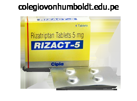
Generic rizact 10mg line
Additionally, evidence has accumulated for an necessary position of a sequence of inflammatory cytokines and chemokines, similar to tumor necrosis factor- and interleukin-1, and progress factors, notably nerve development issue, that are all capable of changing the response properties of pain-signaling neurons. They achieve this in a big selection of methods, including activation or sensitization of nociceptive terminals, as properly as regulation of gene expression by nociceptors. Immune cells are an important source of inflammatory mediators, cytokines, and some development components. Recently, it has become clear that they modulate pain processing not simply by launch of mediators into peripherally damaged or diseased tissue but also by release of the identical mediators into the central nervous system. The newly recognized mediators include quite so much of factors produced and launched from non-neuronal cells, typically immune and glial cells. There is now a rapidly expanding proof base that these are important mediators of persistent pain states and may act at numerous loci. This chapter focuses on the mobile characteristics of nociceptive afferent neurons, their ion channels, and their signal transduction pathways and discusses the ways during which inflammatory mediators impinge on these basic properties. In particular, we first evaluation the cellular mechanisms of activation and sensitization of nociceptors. Then we discuss the roles and actions of particular immune cells and particular ache mediators, beginning with a group of small molecules often rapidly released into damaged tissue. We conclude with a evaluate of the actions of another group of peripheral pain mediators and modulators: the pro-inflammatory cytokines, some chemokines, and some neurotrophic factors, which along with their traditionally recognized roles, are all able to changing the response properties of pain-signaling neurons. That is, they scale back the brink for activation of nociceptors by a number of stimulus modalities and/or improve the responsiveness of nociceptors to suprathreshold stimulation. For a while, attention was focused on a small number of molecules such as prostaglandins and bradykinin. Ion Channels Some inflammatory mediators act by immediately gating the ion channels expressed by sensory neurons. All these ion channels are cation selective and are permeable to either sodium ions or both monovalent and divalent cations. In all instances the ion flow evoked by channel opening depolarizes the sensory neurons and results in neuronal firing. Both lessons of receptors have monomers derived from a single transmembrane phase with a big extracellular ligand-binding area. The useful receptors are both dimers or trimers, which either exist normally or are formed by cross-linking of adjacent monomers by the ligand. In both case, ligand binding prompts kinase pathways that affect gene transcription and can even elicit acute effects on neuronal perform. A, Some of the different stimuli (and the receptors that they act on) that can result in activation and sensitization of the peripheral terminals of nociceptive neurons. B and C show the primary effector mechanisms and second-messenger cascades underlying sensitization, respectively. Bradykinin and the associated peptide kallidin (Lys0-bradykinin) are fashioned from kininogen precursor proteins following the activation of plasma or tissue kallikrein enzymes during inflammation, tissue harm, or anoxia. The biologically active kinins activate two distinct forms of G protein�linked receptors. Bradykinin and kallidin act preferentially at the B2 receptor, whereas des-Arg9-bradykinin and des-Arg10-kallidin act with a lot larger affinity on the B1 receptor than at the B2 receptor. B2 receptors are expressed constitutively on a variety of cell types, including nociceptive sensory nerves, and administration of bradykinin evokes ache and sensitizes polymodal nociceptors (see Mizumura et al 2009). Bradykinin acts immediately on sensory nerves and also can act indirectly by evoking the discharge of different inflammatory mediators from non-neuronal cells. There is good pharmacological evidence that the acute and some of the long-term results of bradykinin are mediated through the B2 receptor. For instance, peptide and non-peptide B2 receptor antagonists have analgesic and anti-hyperalgesic actions in animal fashions of inflammatory ache (Dray and Perkins 1993; Perkins and Kelly 1993, 1994; Asano et al 1997; Burgess et al 2000; Cuhna et al 2007; Valenti et al 2010), in addition to in some neuropathic ache fashions (Werner et al 2007, Luiz et al 2010). There can additionally be immunocytochemical and autoradiographic proof that the B1 receptor is expressed in a subset of sensory neurons (Wotherspoon and Winter 2000, Ma 2001, Petcu et al 2008) and that the level of expression is elevated throughout irritation (Fox et al 2003). Some mediators directly activate cation channels and thus depolarize neurons toward the voltage for initiation of an action potential. Other receptors activate intracellular pathways and influence neuronal sensitivity and excitability indirectly. Phosphorylation or dephosphorylation of membrane proteins often regulates the transduction and transmission of sensory indicators.
Purchase rizact with visa
Top proper: Four microglia surface-rendered with Volocity (Perkin Elmer) software to indicate the adjoining, non-overlapping microdomains of individual cells. Such a spatially restricted system allows the microglial response to react in an anatomically precise style after pathology or harm. The intensive toolbox that microglia possess in terms of cytokines, chemokines, neurotrophins, and neurotransmitters has made this inhabitants of glial cells a wealthy seam of research, and a wealth of data now exists and also stays to be discovered. A common false impression is that resolving these microglial signatures following nerve damage will resolve the behavioral adjustments. Spinal microglia proliferate around the central terminals of peripherally axotomized major afferents. Proliferative and morphological adjustments can be seen by 24 hours after peripheral nerve injury. However, such microglial changes are much less evident in inflammatory and chemotoxic fashions of ache (Honore et al 2000, Clark et al 2007, Lin et al 2007), and the position of microglia within the pain states that result from these insults stays to be elucidated (Li et al 2010). As acknowledged above, microglia exist all through the neuraxis, including at the degree of primary afferents, spinal nociceptive circuitry, and projections to the mind. For a definitive causal function of microglia in pain to be acknowledged, checks of both sufficiency and necessity have to be confirmed. The P2X4R+ microglial phenotype mediates a core pain hypersensitivity cascade following peripheral nerve injury. Chronic neuropathic pain is generated partially by pathological amplification of input to the nociceptive community of the central nervous system. A growing literature has established that neuron� glial interactions inside the spinal wire are responsible, at least in part, for the enhanced output of this network. A specific microglial phenotypic state characterised by up-regulated P2X4R expression (P2X4R+) is induced by peripheral nerve injury and has been shown to play a important position within the pathological modifications in nociceptive processing that underlie neuropathic pain. The lower panel exhibits the advanced modulation of P2X4R expression by varied elements of the parenchymal environment: extracellular matrix (fibronectin; Tsuda et al 2008a, 2009c), infiltrating T cells (Costigan et al 2009a, Tsuda al 2009b), and cytokines and chemokines (Zhang et al 2007, Abbadie et al 2009, Clark et al 2009, Biber et al 2011, Toyomitsu et al 2012). It is thought that the activity of nociceptors is sufficient to elicit a microglial response inasmuch as electrical stimulation of a peripheral nerve at C-fiber depth will induce microglial proliferation in in any other case na�ve animals (Hathway et al 2009). This microgliosis is contiguous with the onset of mechanical hypersensitivity in these animals. However, a extra parsimonious rationalization would counsel a job for A-fiber activity (Suter et al 2009). Corroborating proof for afferent discharge activity driving microglial modifications comes from the observation that the discrete boundaries of microglial proliferation map the anatomical boundaries of the central terminal fields of the injured nerve (Beggs and Salter 2007). Moreover, this improve correlated with the emergence of tactile allodynia (Tsuda et al 2003). Of considerable surprise on the time, it was revealed immunohistochemically that increased expression of P2X4Rs was confined to microglia. An ongoing problem with parsing the actions of P2X subtypes has been the shortage of functional pharmacological instruments (Jarvis and Khakh 2009). Furthermore, as a end result of the behavioral allodynia could be transiently reversed by intrathecal administration of the antagonist, it might be surmised that ongoing P2X4R activation is required to maintain nerve injury�induced allodynia. The demonstration that P2X4R antisense oligonucleotide therapy had an identical action (Tsuda et al 2003) provided additional affirmation. This was definitively proven in experiments during which P2X4Rstimulated microglia had been injected intrathecally into na�ve rats and induced tactile allodynia just like that seen in neuropathic rats (Tsuda et al 2003, Coull et al 2005). The pharmacological, genetic, and behavioral battery of experiments offered the requisite proof to point out sufficiency and necessity of P2X4Rs in mediating neuropathic pain behavior in rats and subsequently logically a causative role. The query then turned to the effectors and effects of P2X4R up-regulation within the improvement and upkeep of neuropathic pain. It is important to notice that these are broad-spectrum agents with antiinflammatory properties. It has been proven that administration of minocycline and propentofylline is simpler in preventing than in reversing nerve injury�induced persistent pain behavior (Raghavendra et al 2003a, 2003b; Ledeboer et al 2005). One interpretation could probably be that microglia have only a transient role in neuropathic ache. An different is that these compounds are lively elsewhere, for example, attenuating the discharge exercise of major afferents following injury (Gong et al 2010). In either case, the clear conclusion is to train caution in attributing faulty mobile specificity to those compounds.
Diseases
- Tinnitus
- Thalassemia major
- Midline developmental field defects
- Partington Mulley syndrome
- Blepharophimosis ptosis esotropia syndactyly short
- Penis agenesia
- Beta-sarcoglycanopathy
- Lubani Al Saleh Teebi syndrome
Buy cheapest rizact and rizact
In addition, within the neck, the vertebral artery gives off spinal branches to the cervical spinal twine and vertebrae and muscular branches. It is important to notice, due to this fact, that variations happen within the origins of the proper subclavian artery, which may come up instantly from the aortic arch either as its first or as its final branch. In the latter case, the right subclavian artery passes behind the trachea and oesophagus in the course to the neck; this vessel could then compress the oesophagus and produce difficulty in swallowing (dysphagia lusoria). Occasionally, the left subclavian artery has a typical origin with the left carotid from the aortic arch. The close relation of the subclavian artery to the brachial plexus accounts for the ache, weak point and numbness within the arm which accompany this lesion. Vascular changes within the arm associated with a cervical rib are most likely as a result of peripheral emboli thrown off from thrombi forming on the partitions of the compressed subclavian artery. The veins of the top and neck the cerebral venous system the venous drainage of the mind follows two pathways: 1 the superficial constructions. The two inside cerebral veins unite to kind the great cerebral vein (the vein of Galen), which emerges from underneath the splenium of the corpus callosum to hitch the inferior sagittal sinus within the formation of the straight sinus. They obtain the venous drainage of the mind and of the skull (the diploic veins) and disgorge 330 the pinnacle and neck Superior sagittal sinus Inferior sagittal sinus Straight sinus Sigmoid sinus Tentorium cerebelli Falx cerebri Great cerebral vein (a) Infundibulum hypophysis cerebri Optic nerve Cavernous sinus Basilar plexus Inferior petrosal sinus Superior petrosal sinus Straight sinus Transverse sinus Internal carotid artery Oculomotor nerve Foramen magnum Transverse sinus Confluence of sinuses (b). They also talk with the veins of the scalp, face and neck by way of emissary veins that move through a variety of the foramina in the skull. The superior sagittal sinus lies along the hooked up edge of the falx cerebri and ends posteriorly (usually) in the right transverse sinus. The inferior sagittal sinus lies within the free margin of the falx cerebri and opens into the straight sinus. The straight sinus lies within the tentorium cerebelli along the attachment of the falx cerebri. It is shaped by the junction of the great cerebral vein of Galen with the inferior sagittal sinus and runs backwards to open (usually) into the left transverse sinus. The transverse sinuses begin at the inside occipital protuberance and run within the tentorium cerebelli on either side alongside its attached margin. On reaching the mastoid a part of the temporal bone each passes downwards, forwards and medially because the sigmoid sinus to emerge via the jugular foramen as the inner jugular vein. Lying above the cavernous sinus are three important structures � the optic tract, the uncus of the temporal lobe of the cerebrum and the internal carotid artery, which first pierces the roof of the sinus then doubles back to lie towards it. The ophthalmic veins drain into the anterior side of the cavernous sinus, which additionally hyperlinks up, by way of these veins, with the pterygoid venous plexus and the anterior facial vein. The cavernous sinus also receives venous drainage from the mind (the superficial middle cerebral vein) and from the dura (the sphenoparietal sinus). Posteriorly, the superior and inferior petrosal sinuses drain the cavernous sinus into the sigmoid sinus and into the commencement of the inner jugular vein, respectively. A attribute image results � blockage of the venous drainage of the orbit causes oedema of the conjunctiva and eyelids and marked exophthalmos, which demonstrates transmitted pulsations from the interior carotid artery. Examination of the fundus exhibits papilloedema, venous engorgement and retinal haemorrhages, all resulting from the acutely obstructed venous drainage. A caroticocavernous arteriovenous fistula outcomes with pulsating exophthalmos, a loud bruit simply heard over the eye and, again, ophthalmoplegia and marked orbital and conjunctival oedema as a outcome of venous stress within the sinus being raised to arterial stage. Close relationship to the mastoid and center ear renders these sinuses liable to infective thrombosis secondary to otitis media. It can also be attainable for sagittal sinus thrombosis to follow infections of the cranium, nostril, face or scalp because of its diploic and emissary vein connections; if there were no emissary veins, infections of the face and scalp would never have achieved their sinister reputation. The internal jugular vein the inner jugular vein runs from its origin on the jugular foramen (as the continuation of the sigmoid sinus) to its termination behind the sternal extremity of the clavicle, the place it joins the subclavian vein to type the brachiocephalic vein. It lies lateral first to the internal after which to the common carotid artery inside the carotid sheath and its relations are subsequently equivalent with these vessels. The deep cervical chain of lymph nodes lies shut against the vein and, if involved by malignant or inflammatory illness, may turn into densely adherent to the vein.

Cheap 5mg rizact with mastercard
Human pain thresholds are generally measured because the temperature that corresponds to the primary report of ache as skin temperature is elevated linearly (Marstock technique). Investigators have noted that faster charges of change in temperature result in lower estimates of the warmth pain threshold (Yarnitsky and Ochoa 1990, Tillman et al 1995a). Warm fibers respond vigorously to gentle warming of the skin and are thought to signal the feeling of heat (Johnson et al 1979). Psychophysical research in people show that pain increases monotonically with stimulus intensities between forty and 50�C. Human Microneurographic Recordings Microneurography has been used to record from nociceptive afferents in awake humans and allows correlations between the discharge of afferents and the reported sensations of the topic. The approach entails percutaneous insertion of a microelectrode into fascicles of nerves such as the superficial radial nerve on the wrist. These studies have demonstrated that the properties of nociceptors in humans and monkeys are comparable. In some experiments the microelectrode can also be used to stimulate an recognized, single nerve fiber in awake human topics to evoke particular sensations. Some, nevertheless, argue that the scale of the stimulating electrode is too large to stimulate particular person models (Wall and McMahon 1985). Correlation of the response of C-fiber nociceptors in the monkey with pain scores in human subjects. The shut match between the curves helps a task for C-fiber nociceptors in warmth ache sensation from glabrous pores and skin. The remaining 9 stimuli ranged from 41�49�C in 1�C increments and have been presented in random order. Human judgments of pain had been measured with a magnitude estimation technique: topics assigned an arbitrary number (the modulus) to the magnitude of ache evoked by the primary 45�C stimulus and judged the painfulness of all subsequent stimuli as a ratio of this modulus. Rather, it appears that nociceptors must attain a certain discharge frequency (about 0. In hairy pores and skin, stepped heat stimuli evoke a double pain sensation (Lewis and Pochin 1937). The first perception is a sharp pricking sensation, and the second sensation is a burning feeling that happens after a momentary lull throughout which little if something is felt. Myelinated afferent fibers should sign the first ache because the latency of response to the first pain is too small to be carried by C fibers (Campbell and LaMotte 1983). The preceding discussion indicates that nociceptors could signal ache in response to warmth stimuli. Central mechanisms, together with attentional and emotional states, quite clearly play a vital function in whether and how much nociceptor exercise results in the notion of ache. For example, the pain in response to mild touch that happens after certain nerve injuries or with tissue harm seems to be signaled by activity in low-threshold mechanoreceptors (see below). When a controlled-force stimulus is utilized to the receptive subject, the response is biggest on the onset of the stimulus after which slowly adapts. Like warmth, repeated shows of a mechanical stimulus lead to pronounced fatigue. Much has been learned concerning the features of a mechanical stimulus that decide the response of nociceptors to mechanical stimuli. The discharge of nociceptors increases with increased pressure and pressure, however these features vary depending on probe measurement: the smaller the probe, the larger the response (Garell et al 1996). For cylindrical probes of various diameter, the discharges are comparable if the intensity of the stimulus is calculated in accordance with force per size of the perimeter of the cylindrical probe. This means that the stress/strain maximum that occurs on the edge of the cylindrical stimulus is the important parameter for excitation of nociceptor terminals. For a given probe dimension, the response of A-fiber nociceptors will increase monotonically with drive, whereas the response of C-fiber nociceptors becomes saturated at greater force levels. Mechanical stimuli had been offered to the receptive field of A-fiber and C-fiber nociceptors at totally different interstimulus intervals (with 10 minutes between stimulus pairs). The A-fiber response (triangles) recovered inside 60 seconds, whereas the C-fiber response (circles) took more than a hundred and fifty seconds to recover. To normalize the information, the response to the test stimulus was divided by the response to the immediately previous conditioning stimulus.
Order 5mg rizact overnight delivery
A successive report of 10 patients399 famous that 4 of 9 patients survived longer than 1 month, and sufferers had a high prevalence of recurrent amyloid deposition in the transplanted coronary heart. A report of 10 patients who received a cardiac transplant between 1984 and 1997 indicated a 20% perioperative mortality rate; the eight surviving sufferers had a 50-month follow-up period. Overall, 7 of the 10 sufferers died after transplantation (median survival, 32 months; vary, 3 to 2113 116 months). The perioperative mortality fee of 20% was attributable to extracardiac amyloid deposits. Heart transplantation is possible technically, however remedy to eliminate the underlying plasma cell proliferative disorder is necessary for a good outcome. Eleven cardiac transplant recipients have received stem cell transplantation at our establishment; of these sufferers, six are alive at 2 to 7 years after the transplantation. Heart transplantation has been reported by numerous teams for the administration of amyloid cardiomyopathy. The United Network for Organ Sharing database contained data of 69 sufferers with a prognosis of amyloidosis who acquired a heart transplant. Patient sex influenced survival: the 1-year survival price was 84% for males and 64% for ladies. Two patients died of progressive amyloidosis at 33 and 90 months after heart transplantation. These survival charges were less than these of sufferers undergoing transplantation for other indications. Progression of amyloidosis contributed substantially to the increased mortality rate. One affected person had a combined heart�liver transplantation, and two patients died after intervention at 23 months. Eight of the sufferers received high-dose chemotherapy adopted by an autologous stem cell transplant. Six of seven evaluable patients achieved a hematologic full remission, and one was a partial remission. At a median follow-up of fifty six months from heart transplant, five of seven sufferers are alive with out recurrence. Their survival was compared with 17,389 sufferers who acquired heart transplant for nonamyloid heart illness: 64% in nonamyloid versus 60% in amyloid patients at 7 years (P =. Seven of eight sufferers who receved a transplant have had no evidence of recurrent amyloid in their cardiac allograft. Angiotensin-converting enzyme inhibitors might reduce proteinuria for sufferers with nephrotic syndrome. Symptoms of toxicity of angiotensin-converting enzyme inhibitors embrace hyperkalemia and hypotension. Enalapril and lisinopril have been reported to decrease proteinuria and corticosteroidresistant nephrotic syndrome that is as a end result of of focal segmental glomerulosclerosis. The possibility that angiotensin-converting enzyme inhibitors could additionally be active in amyloid nephropathy has not been explored. Outcome knowledge were stratified in accordance with three therapy regimens: group 1, kidney transplantation followed by autologous stem cell transplantation (n = 8); group 2, autologous stem cell transplantation followed by kidney transplantation (n = 6); and group three, kidney transplantation after full remission achieved with nonmyeloablative therapy (n = 5) (median followup, 41. Recurrent amyloidosis was recognized through biopsy in a patient in group 2 (before an autologous stem cell transplantation) and in one other patient in group 3. Hemodialysis and chronic ambulatory peritoneal dialysis had equivalent long-term survival rates. Patients mostly succumbed to growth of extrarenal amyloid deposits within the gastrointestinal tract and coronary heart. Patients with kidney amyloid who in the end required dialysis help had considerably greater serum creatinine and 24-hour urine protein levels at prognosis. Patients with l light chains had been significantly extra prone to have kidney involvement and had significantly higher urinary protein loss than these with k mild chain. Serum creatinine degree was an independent predictor of survival even when corrected for heart involvement. For the 38 patients reported who received dialysis, median survival from the initiation of dialysis was 10. Another report described 45 patients with amyloidosis who underwent kidney transplantation. Recurrent amyloid deposits within the allograft had been established histologically for four patients.
Syndromes
- Motor vehicle accidents
- Age 19 and older: 4.7 g/day
- Hump behind shoulders
- This surgery is done through a cut on the left side of the chest, which may reach to the abdomen.
- Chest pain
- Confirm health insurance coverage
Order discount rizact
The second study463 randomly assigned one hundred sufferers to receive colchicine therapy or a combination of melphalan, prednisone, and colchicine. With multivariate evaluation, melphalan had a significant impression on survival when coronary heart failure was not current. Of 153 patients receiving melphalan, cytogenetic abnormalities had been acknowledged in 10. Eight of the 10 sufferers died of pancytopenia, one died of progressive renal amyloidosis, and one was alive at the time of publication. Overall, bone marrow harm consistent with alkylation-induced toxicity was observed for 7% of the patient population. The actuarial risk was 21% for myelodysplasia or acute leukemia developing forty two months after initiation of remedy. The median survival period was eight months after receiving the prognosis of leukemia or myelodysplasia. In one examine, 9 consecutive sufferers were treated with dexamethasone alone (dosage, 40 mg on days 1 via four, days 9 through 12, and days 17 through 20, every 5 weeks for 3 to six cycles). Three patients acquired upkeep ranges of dexamethasone (dosage, 40 mg on days 1 via 4, every month for 1 year). Of seven with nephrotic-range proteinuria, six had a 50% discount in proteinuria, with a median time to response of only four months. Organ operate enchancment was reported in amyloid neuropathy, hepatic involvement, and gastrointestinal involvement. Neither affected person with heart failure as a reduction of more than 30%, with a minimal decrement of 300 pg/ml. Although uncommon, a response in amyloid peripheral neuropathy ought to be documented by an electromyogram exhibiting improved nerve conduction velocities. A consensus definition of organ involvement and remedy response incorporating the free light-chain assay was developed at the Tenth International Symposium on Amyloid and Amyloidosis. The cornerstone of therapy, described in the following section, has been alkylation or corticosteroid based mostly. However, with advances made within the last decade for myeloma remedy, new regimens have been examined, however none have been subjected to randomized scientific trials. In one examine,455 the cytoplasm of the plasma cells contained light-chain tetramers. After successful alkylating agent�based chemotherapy, light-chain synthesis was suppressed and a medical response was noticed. The two earliest reviews of melphalan and prednisone therapy456,457 confirmed responses and prolongation of survival for a minority of patients. Even with subset evaluation, no cohort had a response rate that exceeded 40%, including sufferers with single-organ renal involvement and regular serum creatinine values. Moreover, even when sufferers showed an organ response, follow-up tissue biopsy specimens showed persistent deposits of amyloid. When the Mayo Clinic experience with melphalan and prednisone therapy was reviewed,371 the general response fee was solely 18%. The highest response rate (39%) was noticed with the small group of patients with nephrotic syndrome, no extrarenal involvement, regular serum creatinine values, and normal echocardiographic findings. Responses were noted for sufferers with amyloid cardiomyopathy, suggesting that no affected person is merely too unwell for remedy. Although solely 18% of sufferers responded to melphalan, their median survival time was 89 months. It is commonly tough to know when to discontinue low-dose Chapter ninety nine Immunoglobulin Light-Chain Amyloidosis (Primary Amyloidosis) showed enchancment. Dexamethasone has no tendency to cause leukemia, and the responses are faster than with melphalan. When we handled 19 sufferers with high-dose dexamethasone in an equivalent routine without interferon, only three had an objective organ response. Dexamethasone could additionally be beneficial when treatment with melphalan fails,467 however toxicity may be formidable and should embrace fluid retention, gastrointestinal bleeding, and colonic perforation. The combination of melphalan with high-dose dexamethasone therapy has been reported in the administration of amyloidosis.
Purchase rizact with visa
Instead they occur sequentially in four steps, with an interaction between the subunits occurring in such a means that through the successive combinations of the subunits with oxygen, every combination facilitates the next. Similarly, dissociation of oxygen from hemoglobin subunits facilitates further dissociations. Assuming a blood hemoglobin focus of 15 g Hb/100 mL of blood, this corresponds to fifteen. The affect of pH (and Pco2) on the oxyhemoglobin dissociation curve is referred to as the Bohr effect. High temperatures shift the curve to the proper; low temperatures shift the curve to the left. The Pco2 is larger, the pH is decrease, and the temperature can additionally be larger than that of the arterial blood. In reality, the curve shows that at a Po2 of 70 mm Hg, hemoglobin is still roughly ninety four. This constitutes an essential security factor because a patient with a comparatively low alveolar or arterial Po2 of 70 mm Hg (owing to hypoventilation or intrapulmonary shunt, for example) continues to be in a position to load sufficient oxygen into the blood. A fast calculation exhibits that at 70 mm Hg, the whole blood oxygen content material is roughly 19. This means that a small lower in Po2 may end up in a substantial further dissociation of oxygen and hemoglobin, unloading extra oxygen to be used by the tissues. At a Po2 of forty mm Hg, hemoglobin is about 75% saturated with oxygen, with a total blood oxygen content material of 15. The unloading of oxygen at the tissues can also be facilitated by different physiologic elements that can alter the form and place of the oxyhemoglobin dissociation curve. The P50 is the Po2 at which 50% of the hemoglobin present in the blood is in the deoxyhemoglobin state and 50% is within the oxyhemoglobin state. If the oxyhemoglobin dissociation curve is shifted to the proper, the P50 will increase. It is the quantity of hemoglobin that decreases, not the p.c saturation and even the arterial Po2. Carbon monoxide has a a lot greater affinity for hemoglobin than does oxygen, as mentioned in Chapter 35. Carbon monoxide has a second deleterious effect: it shifts the oxyhemoglobin dissociation curve to the left. It may be attributable to nitrite poisoning or by toxic reactions to oxidant medication, or it can be found congenitally in sufferers with hemoglobin M. As already mentioned on this chapter, variants of the conventional HbA may have totally different affinities for oxygen. Fetal Po2 is way lower than within the grownup; the curve is positioned properly for its operating vary. Myoglobin (Mb), a heme protein that occurs naturally in muscle cells, consists of a single polypeptide chain hooked up to a heme group. It can subsequently combine chemically with a single molecule of oxygen and is comparable structurally to a single subunit of hemoglobin. It can be launched from the Mb when conditions within muscle trigger lower tissue Po2. It is a bluish purple discoloration of the skin, nail beds, and mucous membranes, and its presence is indicative of an abnormally high focus of deoxyhemoglobin in the arterial blood. About 200�250 mL of carbon dioxide is produced by the tissue metabolism each minute in a resting 70-kg individual and must be carried by the venous blood to the lung for elimination from the body. At a cardiac output of 5 L/min, every a hundred mL of blood passing through the lungs must therefore unload 4�5 mL of carbon dioxide. As a outcome, about 5�10% of the whole carbon dioxide transported by the blood is carried in bodily solution. A) the results of carbon monoxide and anemia on the carriage of oxygen by hemoglobin. Note that the ordinate is expressed as the amount of oxygen bound to hemoglobin in milliliters of oxygen per 100 mL of blood. B) A comparison of the oxyhemoglobin dissociation curves for normal adult hemoglobin (HbA) and fetal hemoglobin (HbF). C) Dissociation curves for normal HbA, a single monomeric subunit of hemoglobin (Hb subunit), and myoglobin (Mb). One hundred milliliters of plasma or entire blood at a Pco2 of forty mm Hg, therefore, incorporates about 2.
Buy rizact visa
The affected person achieved a complete hematologic response and was alive 29 months after transplantation. The second patient acquired a stem cell transplant from a human leukocyte antigen�identical sibling after therapy with melphalan (140 mg/m2) and total physique irradiation (800 cGy). The affected person had continual graft-versus-host disease of the skin and liver but was alive 18 months after transplantation. Allogeneic stem cell transplantation may be a promising therapy, however the mortality fee is high. Overall survival and progression-free survival at 1 yr have been 60% and 53%, respectively. For 5 of seven evaluable sufferers who had a complete response, chronic graft-versus-host disease was observed, which implied existence of a graft-versus-amyloidosis impact. It is Chapter 99 Immunoglobulin Light-Chain Amyloidosis (Primary Amyloidosis) and intestinal tract. The median time to onset of bleeding was 9 days after transplantation, and the median platelet rely was 22 � 109/L. Bleeding was seen within the higher gastrointestinal tract for 2 sufferers, the decrease tract for three, and the lower and higher tract for four. Endoscopy was carried out for 5 sufferers, and all had inflamed, friable gastric and esophageal mucosa. Gastrointestinal tract bleeding was related to feminine intercourse and poor engraftment of platelets. During the primary 100 days after transplantation, the median number of packed pink blood cell units administered was 20 for those with gastrointestinal tract bleeding. The exact mechanism underlying bleeding after stem cell transplantation is unknown. The widespread vascular deposits of amyloid may render the vessels inflexible and friable, nevertheless. High-dose chemotherapy, which causes clinically significant mucosal damage, can also cause bleeding. Of patients who received mobilization treatment with cyclophosphamide, the median number of apheresis was three. Of patients receiving filgrastim alone, the median variety of apheresis was two, a statistically significant difference. At Mayo Clinic, 434 patients with amyloidosis obtained their transplants at a median of 4. Predictors of survival included weight achieve of larger than 2% throughout stem cell mobilization and an absolute lymphocyte rely at day 15 of higher than 500 cells/ml. The number of organs involved appeared to be relevant to predicting the outcome efficiently. Patients with two-organ involvement had a median survival fee of 70% at 60 months; and people with three-organ involvement, a median survival of fifty eight months. A neutrophil rely of 500 cells/ml was achieved at a median of 14 days (range, 7 to 116 days). Twentyfive % of sufferers confirmed engraftment on or before day 13, 75% by day 16, and 90% by day 22. A platelet count of 20 � 109/L was achieved at a median of 14 days (range, 6 to 406 days); 25% of patients achieved 20 � 109/L by day 12, 75% by day 19, and 90% by day 27. A platelet count of 50 � 109/L was achieved at a median of 18 days; 25% achieved the depend by day 14, 75% by day 27, and 90% by day 47. Caution is required when interpreting these results, nevertheless; the treatment-related mortality price was excessive (24%). Furthermore, solely 29 of the 100 sufferers who underwent transplantation were evaluable for response. The extent of cardiac amyloidosis measured with echocardiography in the transplant population is lower than that anticipated of an unselected nontransplant cohort. The median intraventricular septal thickness was 12 mm; one quarter of sufferers had a septal thickness of 10 mm or less, and three quarters had less than 14 mm. The absolute lymphocyte depend 15 days after stem cell transplantation may have an impact on survival. In a proportional hazards mannequin, total survival is linked to the pretransplantation free light-chain levels and the variety of organs involved. In giant medical centers that carry out stem cell transplantation, the treatment-related mortality rate is 6% to 18%.
Order rizact from india
In this case, the liver shifts a few of the disposal of ammonium ion from urea to glutamine. Nonrenal mechanisms of acidifying the blood Consumption and metabolism of protein (meat) containing acidic or sulfur-containing amino acids Consumption of acidic medicine Metabolism of substrate with out complete oxidation (fat to ketones and carbohydrate to lactic acid). A decrease in extracellular pH stimulates renal glutamine oxidation by the proximal tubule, whereas a rise does simply the alternative. Thus, an acidosis that lowers plasma pH, by stimulating renal glutamine oxidation, causes the kidneys to contribute extra new bicarbonate to the blood, thereby counteracting the acidosis. Conversely, an alkalosis inhibits glutamine metabolism, resulting in little or no renal contribution of recent bicarbonate via this route. Table 47�3 offers a abstract of the processes of adding acids and bases to the body fluids. The unifying and, subsequently, simplifying principle is that every one processes of acid or base addition boil all the way down to addition or lack of bicarbonate. All processes that acidify the blood find yourself eradicating bicarbonate, and all processes that alkalinize the blood find yourself adding bicarbonate. Compensation exists when both Pco2 or bicarbonate levels stay altered for a time frame and the body modifications the opposite variable in the identical direction. For example, if the Pco2 is abnormally low, renal compensation consists of reducing plasma bicarbonate. Similarly, if bicarbonate is abnormally low, respiratory compensation consists of decreasing arterial Pco2. The compensatory modifications convey the ratio of bicarbonate to Pco2 closer to a traditional value, and therefore decrease the change in pH. Consider a case where the Pco2 is simply too excessive (respiratory acidosis) as a outcome of hypoventilation (review Chapter 37). The astute reader might recognize a possible downside within the interpretation of acid�base issues. Is this a respiratory acidosis with renal compensation, or is it a metabolic alkalosis with respiratory compensation Fortunately in a clinical setting it would be uncommon to not have additional info. For instance, the excessive Pco2 of persistent bronchitis affected person is, in all likelihood, a respiratory acidosis resulting from impaired ventilation, not a compensation for a metabolic alkalosis. Nevertheless, in real life there are often mixed acid�base problems that indeed current a problem within the clinic. This subject is roofed extra totally within the context of the respiratory system in Chapter 37. Acid�base issues develop when both arterial Pco2, bicarbonate, or each deviate from their normal vary. The pH could be restored to regular if the bicarbonate have been elevated to the same degree as Paco2. In any metabolic alkalosis, by definition the plasma bicarbonate concentration is elevated. The decreased Pco2 and improve in extracellular pH signal reduced tubular hydrogen ion secretion and elevated bicarbonate secretion. Bicarbonate is misplaced from the body, and the loss ends in decreased plasma bicarbonate and a return of plasma pH toward normal. Besides stimulating sodium reabsorption, aldosterone stimulates hydrogen ion secretion by kind A intercalated cells. The urine, as an alternative of being alkaline, as it should be when the kidneys are usually responding to a metabolic alkalosis, is considerably acid. The generation or upkeep of a metabolic alkalosis in quantity contraction may occur when the amount is normal or high but the body "thinks" volume is low, particularly in congestive heart failure and advanced liver cirrhosis. These embrace (1) elevated input of acid by ingestion, infusion, or manufacturing; (2) decreased renal manufacturing of bicarbonate, as in renal failure; or (3) direct lack of bicarbonate from the body, as in diarrhea. This is precisely what healthy kidneys do in the case of any acid load, but if the acid load is simply too great or the problem is in the kidneys themselves, the bicarbonate focus will remain low. However, we emphasize that specific chloride depletion, in a manner independent of and in addition to extracellular volume contraction, helps keep metabolic alkalosis by stimulating hydrogen ion secretion. The most typical reasons for chloride depletion are persistent vomiting and heavy use of diuretics. However, in some situations, the kidneys fail to do this, and thereby both generate a metabolic alkalosis or keep a metabolic alkalosis that originates from one other cause. Recall that secretion of hydrogen ions, after all filtered bicarbonate has been reabsorbed, generates new bicarbonate.
Order rizact 5mg
The line of the carotid sheath may be marked out by a line becoming a member of a point midway between the tip of the mastoid course of and the angle of the jaw to the sternoclavicular joint. Along this line, the common carotid bifurcates into the external and internal carotid arteries on the stage of the upper border of the thyroid cartilage; at this stage, the vessels lie within the carotid sheath just beneath the investing layer of the deep cervical fascia, the place their pulsation is palpable and sometimes visible. Tissue planes and fascial layers within the anterior a part of the neck Deep to the pores and skin of the neck is the superficial fascia or panniculus adiposus, which is actually a layer of subcutaneous fat, kind of homogeneous. The diploma of adiposity in this layer varies between people; it additionally varies, to some extent, between the anterior and posterior aspects of the neck in the same individual, being typically considerably thinner in the entrance of the neck than in the again. Lying instantly deep to the subcutaneous fat, on both aspect of the anterior midline, is the platysma, a relatively skinny but wide sheet of muscle. The deep fascia can be categorised into four elements: investing layer of deep cervical fascia, pretracheal fascia, prevertebral fascia and carotid sheaths (right and left). Immediately deep to the platysma is the investing layer of deep cervical fascia, essentially the most superficial of the a quantity of layers of the deep cervical fascia. Superiorly, its attachment could additionally be traced circumferentially alongside the complete size of the decrease border of the mandible, the mastoid processes and superior nuchal strains on both facet and to the exterior occipital protuberance within the posterior midline. In the interval between the angle of the mandible and the mastoid process, the investing layer of deep cervical fascia encloses the parotid salivary gland because the parotid fascia. Inferiorly, the circumferential attachment of the investing layer of deep cervical fascia is to the sternal notch. Traced laterally from the anterior midline, between its upper and lower attachments, the investing layer of deep cervical fascia meets, on all sides, the medial border of the cor- the floor anatomy of the neck 289 responding sternocleidomastoid muscle and splits to surround the muscle. Thereafter, it continues posterolaterally as the fascial roof of the posterior triangle of the neck, and, upon reaching the anterior edge of the trapezius muscle, it splits to enclose the trapezius. In its descent from the lower border of the mandible, the investing layer of deep cervical fascia is firmly adherent to the entrance of the hyoid body and to the lateral aspects of the larger horns of the hyoid. Thus, all of the cervical viscera, major blood vessels and nerves of the neck and all of the cervical muscles (with the only real exception of the platysma) come to lie within the sweep of the investing layer of deep cervical fascia. The exterior jugular vein runs in the aircraft between the platysma and the underlying investing layer of deep fascia. Lying immediately deep to the investing layer of deep cervical fascia and operating longitudinally on either facet of the anterior midline of the neck are the infrahyoid anterior cervical muscles, also referred to as the strap muscular tissues. On both sides of the vertical midline, the strap muscular tissues are disposed in two planes. The superficial plane consists of the sternohyoid and omohyoid muscles mendacity facet by facet (sternohyoid medial to omohyoid), and the deep plane consists of the sternothyroid muscle, which extends vertically from the posterior floor of the manubrium sterni to the oblique line of the thyroid cartilage. Extending upwards from the oblique line of the thyroid cartilage to the greater horn of the hyoid is the thyrohyoid muscle, typically thought to be the upward continuation of the sternothyroid muscle. The deepest layer of the deep cervical fascia is the prevertebral fascia, a comparatively dense layer that covers the anterior elements of the prevertebral musculature and the cervical vertebral column. The prevertebral fascia passes across the vertebrae and prevertebral muscle tissue behind the oesophagus, the pharynx and the nice vessels. Laterally, the fascia covers the scalene muscles along with the phrenic nerve, as this lies on scalenus anterior, and the rising brachial plexus and subclavian artery. These buildings carry with them a sheath formed from the prevertebral fascia, which turns into the axillary sheath. Inferiorly, the fascia blends with the anterior longitudinal ligament of the higher thoracic vertebrae in the posterior mediastinum. Pus from a tuberculous cervical vertebra bulges behind this dense fascial layer and will form a midline swelling in the posterior wall of the pharynx. The abscess might then track laterally, deep to the prevertebral fascia, to some extent behind the sternocleidomastoid. Deep to the strap muscles, and anterior to the prevertebral fascial layer, is the centrally located visceral compartment of the neck. Lying lateral to the cervical visceral column, and in front of the prevertebral fascia, are the best and left carotid sheaths. Situated posteromedial 290 the head and neck to every carotid sheath and anterior to the prevertebral fascia is the ganglionated, cervical sympathetic chain. The cervical visceral compartment flanked by the right and left carotid sheaths comprises, most posteriorly, the pharynx and its distal continuation � the oesophagus.
Real Experiences: Customer Reviews on Rizact
Ben, 24 years: A urine dipstick check reveals a light proteinuria (increased protein within the urine), and samples of his blood and urine are despatched to the scientific laboratory for a Reabsorption within the proximal tubule is iso-osmotic. The musculocutaneous nerve the musculocutaneous nerve (C5, C6, C7) continues on from the lateral twine of the plexus. High-risk consists of deletion 17p, t(14;16), t(14;20), but with this new system, sufferers were reclassified into a model new high-risk group, transferring these sufferers previously classified as high-risk however with a trisomy into the standard-risk group. Nociceptive Neurons Defined by their excitatory enter, nociceptive neurons include all neurons which are excited by noxious stimuli.
Bufford, 45 years: The membranes of the brain and spinal cord (the meninges) Three concentrically organized membranes, often identified as meningeal layers or meninges, encompass the brain and spinal wire. In the patients who had demonstrated a previous response (and subsequently progressed) to fludarabine, however, three of four sufferers responded. The perineal physique (central perineal tendon) this fibromuscular node lies in the midline on the junction of the anterior and posterior perineum. However, such microglial changes are much less evident in inflammatory and chemotoxic fashions of ache (Honore et al 2000, Clark et al 2007, Lin et al 2007), and the role of microglia in the ache states that result from these insults stays to be elucidated (Li et al 2010).
8 of 10 - Review by B. Kasim
Votes: 320 votes
Total customer reviews: 320
References
- Rauch A, Dorr HG. Chromosome 5q subtelomeric deletion syndrome. Am J Med Genet C Semin Med Genet. 2007; 145C:372-76.
- Pak CY, Sakhaee K, Pearle MS: Detection of absorptive hypercalciuria type I without the oral calcium load test, J Urol 185(3):915n919, 2011.
- Schlaich MP, Sobotka PA, Krum H, Lambert E, Esler MD. Renal sympathetic-nerve ablation for uncontrolled hypertension. N Engl J Med 2009;361:932-934.
- Li WQ, Ma JL, Zhang L, et al. Effects of Helicobacter pylori treatment on gastric cancer incidence and mortality in subgroups. J Natl Cancer Inst 2014;106(7):dju116.
- Perez EA. Microtubule inhibitors: differentiating tubulin-inhibiting agents based on mechanisms of action, clinical activity, and resistance. Mol Cancer Ther 2009;8(8):2086-2095.


