


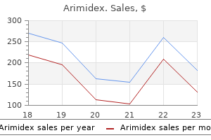
Arimidex
Arimidex dosages: 1 mg
Arimidex packs: 30 pills, 60 pills, 90 pills

Generic arimidex 1 mg with visa
During postnatal improvement, tendons enlarge by interstitial growth, significantly at myotendinous junctions, where there are excessive concentrations of fibroblasts. The thickness lastly attained by a tendon is dependent upon the size and strength of the associated muscle, however it also seems to be influenced by different elements, such because the degree of pennation of the muscle. The metabolic rate of tendons could be very low but increases throughout an infection or injury. Repair entails the preliminary proliferation of fibroblasts, followed by interstitial deposition of new fibres. Specialized endings which would possibly be sensitive to drive (Golgi tendon organs) are discovered close to myotendinous junctions; their giant myelinated afferent axons run centrally within branches of muscular nerves or in small rami of adjoining peripheral nerves. Muscles such as these of the abdominal wall, which originate from a quantity of embryonic segments, are provided by a couple of nerve. In most cases, the nerve travels with the principal blood vessels within a neurovascular bundle, approaches the muscle near its least mobile attachment and enters the deep surface at a position that is sort of constant for each muscle. The motor component consists mainly of huge, myelinated -efferent axons that provide the muscle fibres, supplemented by small, thinly myelinated -efferents, or fusimotor fibres, that innervate the intrafusal muscle fibres of neuromuscular spindles and fantastic, non-myelinated autonomic efferents (C fibres) that innervate vascular clean muscle. Within muscles, nerves observe the connective tissue sheaths, coursing in the epimysial and perimysial septa earlier than getting into the fantastic endomysial tissue across the muscle fibres. The motor axons department repeatedly earlier than they lose their myelinated sheaths and terminate near the center of muscle fibres. These terminals tend to cluster in a slender zone towards the centre of the muscle belly known as the motor point. The axon terminal gives off a quantity of quick, tortuous branches, every ending in an elliptical area, the motor end-plate. Within the underlying discoidal patch of sarcolemma-the sole plate or subneural apparatus-the sarcolemma is thrown into deep synaptic folds. Mutation leads to the absence of this protein in skeletal muscle fibres (as properly as in heart, brain and easy muscle). The dystrophin gene may be very massive, and a number of gene mutations can end result in the defect. Some mutations end in incomplete lack of dystrophin and milder scientific illness. Within muscle, dystrophin is found within the periphery of the fibres, near the cytoplasmic face of the sarcolemma. It is part of a large complex of glycoproteins that spans the muscle membrane, binding intracellular actin with merosin within the muscle basal lamina, thus stabilizing the sarcolemmal membrane and permitting the transmission of forces of contraction and rest from internally to the extracellular matrix. Without dystrophin, the muscle membrane may be extra vulnerable to disruption, with destruction of muscle fibres and growing fibrosis. His father and paternal uncle each skilled sudden cardiac demise of their 50s; though neither had significant muscle weak point, his father had cataracts removed in his 30s. Examination demonstrates facial weakness (diplegia), with delicate bilateral eyelid ptosis and marked frontal balding. He has bilateral distal upper extremity weak point and both proximal and distal lower extremity weak spot. Delayed rest of grip is hanging (myotonia), and percussion of the thenar eminence documents delayed relaxation. Discussion: this man has typical myotonic dystrophy, an autosomal dominant disorder with variable expression. The myotonic phenomenon exhibited on each physical examination and electrophysiological testing is because of repetitive spontaneous and contraction-induced muscle fibre activation. Muscle biopsy reveals a combination of findings, together with muscle fibre atrophy and hypertrophy, some necrotic fibres, fibrosis and adipose deposition, with rare eosinophilic cytoplasmic inclusions. Ringbinden (aberrant muscle fibres encircling usually oriented fibres) could also be found. It is noteworthy that whereas muscular involvement is proximal in the great majority of muscular dystrophies, in myotonic dystrophy a distal distribution is attribute.
Purchase cheap arimidex
These tributaries may exhibit reflux and may be an alternate source of varicosities. In this explicit patient, the incompetent tributary vein is the dominant etiology for the varicose veins. Currently obtainable thermal ablation expertise requires a relatively straight course to the goal vein for gadget passage. Azene E et al: Foamed bleomycin sclerosis of airway venous malformations: the function of interspecialty collaboration. A coaxial microcatheter was placed into the feeding artery, and Onyx embolization was performed. A fluoroscopic image after embolization reveals an intensive Onyx solid and its relationship to the coils and the ureteral stent. Venous Malformation Sclerotherapy (Direct Percutaneous Venography) Venous Malformation Sclerotherapy (Injection of Sclerosant) (Left) After tightly applying a tourniquet above the malformation, a mix of contrast and 3% Sotradecol was slowly launched by way of the angiocatheter and was allowed to stay indwelling for five minutes. The volume needed to fully opacify the malformation by venography was used to calculate the suitable remedy quantity. Absence of shade signal within the mass is due to very sluggish flow in this combined lymphatic-venous malformation. Contrast injection fills the vascular channels and shows a small communication to the ulnar artery. After inserting a tourniquet distally, this mixture was slowly injected into the malformation. Fluoroscopic monitoring was used during injection to confirm complete filling of the malformation and no nontarget reflux. Lymphatic Malformations (Classification by Morphology) Lymphatic Malformations (Lymphangiography of Lymphocele) (Left) Lymphatic malformations are sponge-like collections of abnormal lymphatic channels &/or areas. Physical examination revealed a waterhammer pulse at the anastomosis and an simply compressible main physique of the fistula, findings consistent with juxtaanastomotic stenosis. Retrograde puncture was carried out, and preliminary angiogram was done while occluding the outflow, confirming the suspected stenosis. Repeat bodily exam showed extra turgor within the physique of the fistula and backbone of the abnormal pulsatility at the anastomosis. Outflow Stenosis Problem Detected During cannulation Findings on palpation Findings on auscultation Flow price Static venous pressures Inflow Stenosis Outflow Stenosis Difficulty with cannulation (lacks turgor for easy Prolonged bleeding (> 10 min) after needle puncture) withdrawal Weak thrill Weak bruit Decreased Normal or low Markedly pulsatile Discontinuous bruit; high pitched or focal at web site of stenosis Decreased Increased Objective Measures Triggering Hemodialysis Access Evaluation Measurement Kt/V (dialysis adequacy) Static venous pressures Recirculation Access move rates 1 = imply entry pressure/mean arterial stress. Some proceduralists use ultrasoundguidance during access of deep or doubtlessly thrombosed grafts and fistula. Mild stenosis at the venous anastomosis and an adjacent venous aneurysm is current. Additional imaging of the venous outflow can additionally be obtained by way of this catheter. The guidewire facilitates placement of a vascular access sheath, which may then be used to introduce any catheters which may be wanted for intervention. This patient was referred for ultrasound after which intervention due to low Kt/V. Initial injection confirmed the suspicion of narrowing just downstream from the graft to vein anastomosis. A balloon was positioned throughout the positioning of extravasation and inflated to three atm of pressure. Risk factors for anastomotic disruption include a just lately created access (< 1 month) or inappropriate balloon oversizing. The initial ultrasound revealed a comparatively long-segment stenosis of the juxtaanastomotic region attributable to exuberant venous neointimal hyperplasia. A fistulogram obtained via retrograde entry exhibits the lengthy phase juxtaanastomotic narrowing. A 4-Fr catheter was advanced retrograde via the fistula into the upstream brachial artery.
Arimidex 1mg cheap
Neoplasms of mesenchymal origin, corresponding to osteosarcomas and chondrosarcomas, as properly as malignant lymphomas are much much less frequent. Metastases from other malignancies are occasionally found, with the primary tumor residing within the kidney, lung, breast, testis, or thyroid gland. The main sites of predilection are the nasal cavity and maxillary sinus, adopted by the ethmoid cells, frontal sinus, and sphenoid sinus. Symptoms that are suspicious for malignancy include sudden onset of obstructed nasal breathing combined with bloody rhinorrhea and a fetid nasal odor, particularly in patients over 50 years of age. Advanced tumor phases could also be marked by swelling of the buccal delicate tissues, swelling on the medial canthus of the eye, headache, facial pain, and hypoesthesia or numbness of the cheek as a end result of infraorbital nerve involvement. Orbital infiltration can result in displacement of the orbital contents, diplopia, or proptosis. Osteomas Osteomas are benign bone tumors which will occur as isolated lots, especially within the ethmoid cells and frontal sinus, or may form in depth masses that develop alongside the skull base. Symptoms and analysis: Many of those tumors are detected by the way on x-ray films of the cranium. Computed Probst-Grevers-Iro, Basic Otorhinolaryngology� 2006 Thieme All rights reserved. Diagnosis: the clinical examination contains endoscopic inspection of the nasal cavity. Since sinus tumors are apt to invade the nasal cavity secondarily, endoscopy alone might provide little information on the extent of the mass. For this reason, computed tomography and/or magnetic resonance imaging ought to all the time be performed. Treatment: Treatment is individualized based on the histology and extent of the malignant tumor, and the therapy plan must be coordinated with the radiotherapist and medical oncologist. Since the great majority of lesions are squamous cell carcinomas, however, the remedy of selection will usually encompass surgery and postoperative radiation. The objective of radical tumor elimination could require a very intensive process with partial or full removing of the maxilla or partial resection of the anterior skull base. As a results of shut interdisciplinary cooperation with neurosurgeons, maxillofacial surgeons and ophthalmologists, as nicely as trendy intensive-care options, even very extensive sinonasal malignancies can now be managed by surgical therapy. Since only about 20% of sinonasal malignancies metastasize to regional lymph nodes, a neck dis-. Clinically, esthesioneuroblastoma stays asymptomatic for a while because of its location in the olfactory groove between the upper portions of the nasal septum and the attachment of the middle turbinate. When advanced, the tumor causes obstructed nasal breathing, recurrent epistaxis, and notably hyposmia or anosmia. Some of those tumors turn out to be symptomatic only after invading the cranial cavity or orbit, inflicting headache or visible deterioration. In a few cases, cervical lymph-node metastases are the first manifestation of the disease. Diagnosis relies on endoscopy (see picture below) and particularly computed tomography or magnetic resonance imaging; only these modalities can precisely define the tumor extent. Treatment relies on a mixture of tumor resection and postoperative radiotherapy. Finally, a large share of the taste receptors are located in the oral cavity. The lips and the soft tissues of the cheek function as the outer boundary of the oral vestibule and oral cavity, which form the preliminary part of the digestive tract. The tongue is located such that the body of the tongue is inside the oral cavity, whereas the bottom (root) of the tongue is within the oropharynx, forming its anterior boundary. For studying purposes, however, the tongue as an entire is included within the chapter on the oral cavity. Anatomy Oral Vestibule the oral vestibule is bounded externally by the lips and cheeks and internally by the alveolar processes and teeth. When the teeth are in occlusion, the oral vestibule communicates with the oral cavity via an area behind the last molar. Knowledge about the lymphatic drainage of the lips is essential for understanding the lymphogenous metastasis of malignant tumors of the lips.
Order arimidex 1mg without a prescription
Deep rami communicantes from this plexus be part of the Preganglionic parasympathetic neurone cell our bodies are situated in certain cranial nerve nuclei of the brain stem. In the cranial part of the parasympathetic system there are 4 small peripheral ganglia-ciliary, pterygopalatine, submandibular and otic-which are all described on a regional foundation. These are solely efferent parasympathetic ganglia, not like the trigeminal, facial, glossopharyngeal and vagal ganglia, all of which are concerned exclusively with afferent impulses and comprise the cell bodies of sensory neurones. However, the cranial parasympathetic ganglia are traversed by afferent fibres, postganglionic sympathetic fibres and, within the case of the otic ganglion, branchial efferent fibres, however none of those are interrupted in the ganglia. Postganglionic parasympathetic fibres are often non-myelinated and shorter than those in the sympathetic system, as a end result of the ganglia by which the previous synapse are in or near the viscera they provide. In distinction to the sympathetic system, postganglionic parasympathetic neurones are cholinergic. Oculomotor preganglionic parasympathetic fibres originate in the Edinger� Westphal nucleus of the midbrain and travel within the nerve alongside its department to the inferior indirect, reaching the ciliary ganglion, where they synapse. Postganglionic fibres, that are thinly myelinated, travel in the brief ciliary nerves that pierce the sclera to run ahead within the perichoroidal area to the 371 Chapter 21 Transverse process, C. It has been suggested that the lesion includes preganglionic fibres originating at T2 and T3. Occasionally, segmental autonomic dysfunction is noticed in an higher extremity as properly. A relationship to Ross syndrome (segmental anhidrosis, tonic pupils and hyporeflexia) has additionally been suggested. A 55-year-old girl presents with a 3-month history of exercise-induced anhidrosis and lack of flushing on the left side of the face, with regular autonomic responses on the best. Provocative manoeuvres similar to managed train verify lack of flushing on the left facet of the face, as described historically, along with hemifacial anhidrosis. Discussion: Asymmetric facial flushing and sweating recommend a prognosis of harlequin syndrome, with uneven involvement of vasomotor and sudomotor fibres. Their activation mediates accommodation of the attention to near objects and pupillary constriction. The facial nerve contains preganglionic parasympathetic axons of neurones with their somata within the superior salivatory nucleus (see Ch. The fibres emerge from the mind stem within the nervus intermedius, go away the main facial nerve trunk above the stylomastoid foramen and travel within the chorda tympani, which subsequently joins the lingual nerve (see Ch. In this way, preganglionic fibres are conveyed to the submandibular ganglion, the place they synapse on ganglionic neurones. Postganglionic fibres innervate the submandibular and sublingual salivary glands and are stated to travel in the lingual nerve. Some preganglionic fibres could synapse round cells within the hilum of the submandibular gland. Stimulation of the chorda tympani dilates the arterioles in both glands, in addition to having a direct secretomotor effect. The facial nerve additionally incorporates efferent parasympathetic lacrimal secretomotor axons, which journey in its greater petrosal branch and then through the nerve of the pterygoid canal, to relay within the pterygopalatine ganglion. Postganglionic axons are thought to travel by the zygomatic nerve to the lacrimal gland and by ganglionic branches to the nasal and palatal glands. The glossopharyngeal nerve accommodates preganglionic parasympathetic secretomotor fibres for the parotid gland. These originate in the inferior salivatory nucleus and travel within the glossopharyngeal nerve and its tympanic department. Magnetic resonance imaging reveals a tumour involving the dorsal midbrain (collicular plate) that can additionally be answerable for obstructive hydrocephalus. Lightnear dissociation (characterized by a poor pupillary response (reflex) to gentle, however with preservation of pupillary constriction to a close to target) often results from bilateral midbrain lesions, however not necessarily. It is of interest that Argyll Robertson pupils, which are seen, for example, in circumstances of neurosyphilis, can also exhibit pupillary-near dissociation, but the Argyll Robertson pupil is often very small and irregular, with lowered dilatation in the dark. Again, the supranuclear connection between the pretectum and the midbrain Edinger�Westphal nucleus is spared, so that the pupillarynear reflex is preserved. However, many peripheral ganglia comprise circuits that are capable of sustaining and modulating visceral actions by native reflex mechanisms. Large populations of intrinsic neurones exist which are derived from the neural crest and are impartial of sympathetic and parasympathetic nerves.

Generic arimidex 1 mg amex
Often azathioprine can additionally be prescribed and the dose is adjusted according to the white cell rely. Patients are sometimes nicely informed about symptoms suggesting rejection � usually these resemble an assault of pericarditis. The patient could know of boosts of prednisone which were given for rejection episodes. Later rejection tends to be milder and should reply to a rise in oral steroids. Repetitive rejection could also be handled with total lymphoid irradiation or methotrexate. Many patients are also taking common antibiotics to stop Pneumocystis jirovecii (formerly carinii) an infection. Note the small scars within the neck on the point of introduction of the endomyocardial biopsy forceps. Examine the chest rigorously for indicators of infection, study the mouth for candidiasis, search for an infection at intravenous entry sites and look at the temperature chart. Investigations these depend somewhat on the explanation the patient has been admitted to hospital on this event. If the affected person is presently an inpatient, find out why he or she has been admitted to hospital on this event. Coronary artery intimal proliferation may cause ischaemic coronary heart illness within the transplanted coronary heart. However, there at the moment are patients in whom re-innervation seems to have occurred and led to symptoms of angina. This allograft arteriopathy is among the most important problems after transplant. The situation is usually diffuse, but as soon as lesions causing 40% coronary stenosis have occurred, the prognosis is quite poor: the 2-year survival fee is only about 50%. The situation is current in 10% of recipients at 1-year post-transplant and in 50% at 5 years. Find out whether or not the patient knows what his or her ldl cholesterol stage is and what therapy is getting used to hold it low. Intravascular ultrasound is used more and more to detect subclinical vasculopathy and to study the benefits of various antirejection regimens. The incidence in heart transplant patients is lower than that for these with liver transplants, but greater than that for these with renal transplants. Reduction in the amount of immunosuppression will typically assist, but antiviral treatment with acyclovir or interferon could additionally be needed. The usual approach is to give the patient a lift of methylprednisolone � usually 1 g intravenously day by day for three days, adopted by a repeat biopsy. Possible episodes of infection should be investigated completely and treated aggressively with appropriate remedy. Further cardiac failure may be an indication of rejection, which should be treated. Hyperlipidaemia ought to be sought routinely and treated most vigorously with medicine and food regimen. The 1-year survival fee after transplant is about 90% and the 5-year survival rate is about 75%. The dialogue of a affected person awaiting cardiac transplantation will probably run along related strains. Some sufferers awaiting transplantation are sick sufficient to require intravenous inotropes. There may even have been speak about the utilization of exterior circulatory assistance gadgets for use as a bridge to transplant. Chest X-ray may show signs of cardiac enlargement, though this is a late sign of rejection.
Cotton. Arimidex.
- What is Cotton?
- Are there safety concerns?
- Dosing considerations for Cotton.
- How does Cotton work?
- Menstrual disorders, menopausal symptoms, nausea, fever, headache, diarrhea, kidney and bladder conditions, inducing labor and delivery, male contraception, and other conditions.
Source: http://www.rxlist.com/script/main/art.asp?articlekey=96428
Order arimidex 1 mg otc
This permits Huber needles to repeatedly penetrate the silicon cap of a port without coring or cutting the silicone. Coady K et al: A comparison of infections and complications in central venous catheters in adults with solid tumours. Chest Port Insertion, Step-by-Step (Subcutaneous Pocket Creation) Chest Port Insertion, Step-by-Step (Subcutaneous Tunnel) (Left) the microwire is replaced with an zero. Then, an incision of sufficient length to accommodate the port is made in the anterior chest wall, 2-3 fingers width beneath the clavicle. Blunt dissection with a hemostat creates a subcutaneous pocket just giant sufficient for the port. The catheter must be instantly adjoining to the peel-away sheath with no intervening skin. Skin closure is achieved with an absorbable running subcuticular stitch, or simply with "glue" and Steri-Strips. Chest Port Insertion, Step-by-Step (Subcutaneous Pocket Closure) Catheter Tip Position (Patient Supine) (Left) Following port placement, imaging must be obtained to doc port position and exclude problems. Port catheter tip location, nevertheless, can change dramatically as seen on this patient imaged supine. Especially in obese or largebreasted ladies, the port is pulled down and the catheter tip up when the affected person is upright. This motion can be minimized by placing the port higher, closer to the clavicle. Catheter Tip Position (Patient Upright) Chest Port Complication (Hematoma) (Left) A patient on chronic anticoagulation therapy has intensive ecchymosis overlying the port pocket, extending along the subcutaneous catheter tunnel. This resolving hematoma is the most common complication associated with port insertion. Administration of the angiogenesis inhibitor bevacizumab inside 2 weeks earlier than or after insertion will increase the chance of this complication. Fluoroscopic imaging confirms that the Huber needle is nicely positioned within the reservoir. Two (optional) 3-0 Vicryl stay sutures had been utilized for port reservoir attachment. Arm Port Placement, Step-by-Step (Subcutaneous Pocket Creation) one hundred thirty Ports Venous, Portal, and Lymphatic Procedures Arm Port Placement, Step-by-Step (Catheter Tunnel and Attachment) Arm Port Placement, Step-by-Step (Fluoroscopic Imaging) (Left) the catheter is now tunneled kind the port pocket via the subcutaneous airplane to the venotomy web site. The catheter is then detached from the tunneler and superior through to the peelaway sheath to the best atrium. Lumbar Port Placement (Final Port Position) Lumbar Port Complication (Partial Inferior Vena Cava Thrombosis) (Left) A subcutaneous pocket is created overlying a lower rib or the iliac crest to present underlying support. The subclavian vein programs anterior to the subclavian artery, the anterior scalene muscle, and the 1st rib, and is posterior to the clavicle. This young male affected person had acute onset of arm swelling, typical of venous thoracic outlet syndrome. Primary Axillosubclavian Vein Thrombosis Central Venous Occlusion (Left) A central venogram was obtained via a long right brachial vein sheath on this 77-year-old lady requiring new access for hemodialysis. Paget-Schroetter syndrome, effort thrombosis, venous thoracic outlet syndrome � Thrombosis/stenosis/occlusion of axillary/subclavian vein � Occurs in younger adults following repetitive workout routines. Breault S et al: Percutaneous endovascular administration of continual superior vena cava syndrome of benign causes: long-term follow-up. The arm pain and swelling that occurs with acute thrombosis often improves as venous collaterals shortly develop. In-stent restenosis generally occurs with narrowing often located on the costoclavicular junction. No stent was necessary since there was no residual narrowing on subsequent venography. Occluded Right Brachiocephalic Vein (Oblique View During Recanalization) 142 Upper Extremity and Central Venous Intervention Venous, Portal, and Lymphatic Procedures Occluded Left Brachiocephalic Vein (Diagnostic Venogram) Occluded Left Brachiocephalic Vein (Post Stent Placement) (Left) A left arm hemodialysis fistulogram revealed occlusion of the left brachiocephalic vein and enlarged draining venous collaterals in the right neck. Venous drainage from the left arm is markedly improved, and the collaterals are now not distinguished. Therefore, a stent was placed throughout the left brachiocephalic stenosis, whereas a parallel stent was placed into the azygos vein to keep patency of that venous drainage route. There is marked distension of the brachiocephalic, subclavian, and axillary veins, in addition to of the external and inside jugular veins. Contrast was concurrently injected by way of the best internal jugular sheath and through the catheter so as to fill vessels above and under the obstruction.
Buy genuine arimidex line
Note the contralateral high-attenuation left renal cyst (confirmed at ultrasound). Note the delicate wall calcification and a nephrogram delay, which is likely due to a mixture of shunting and mass effect upon a partially obstructed renal pelvis. This is a uncommon but extremely aggressive tumor that impacts young males with sickle cell trait and has a really poor prognosis. Common and uncommon histologic subtypes of renal cell carcinoma: imaging spectrum with pathologic correlation. Features of these Bosniak I cysts embrace water attenuation, the dearth of an enhancing wall, septa, or calcifications. Ultrasound is an adjunct modality in Bosniak classification system and is a helpful method to differentiate between high-attenuation cysts and true solid renal lesions. Benign renal cysts are present in 20-30% of middle-aged adults, and the incidence will increase with age. Cyst septation is accentuated at ultrasound and sometimes erroneously prompts Bosniak upclassification. Care should be taken to differentiate noncommunicating peripelvic cysts from hydronephrosis at ultrasound. Complexity and perinephric infiltration recommend an abscess, however scientific historical past, urinalysis, and aspiration affirm the diagnosis. The characteristic imaging appearance of this acquired (not hereditary) entity is a nonencapsulated collection of cysts. Partial nephrectomy confirmed a combined epithelial and stromal tumor, a uncommon benign cystic renal neoplasm. Diagnosis and administration of sufferers with difficult cystic lesions of the kidney. Gastric wall is thickened as a end result of gastritis induced by immunosuppressive medicine on this affected person who had a renal transplant following years of dialysis therapy. Note the infiltrating renal cell carcinoma with tumor enlargement of the best renal vein. The hypervascularity of the mass and the invasion of the renal vein counsel the diagnosis of renal cell carcinoma, rather than transitional cell carcinoma. Low-density xanthomatous irritation replaces the renal parenchyma and spreads to the perirenal house. In the proper kidney, there are rounded parenchymal defects, while the left kidney reveals more wedgeshaped lesions, resembling acute pyelonephritis. Note the heterogeneous fat density mass, an acute myeloid leukemia, that was the source of the hemorrhage. The left ureter had been injured during a colonic resection, causing a stricture and urinary leak. These findings plus pleural thickening and sclerosis of the metaphyseal regions of the femur are attribute options of Erdheim-Chester disease. Biopsy of lymph nodes confirmed sinus histiocytosis (Rosai-Dorfman disease) that accounted for all findings, including the perirenal infiltration. The constellation of findings (pleural thickening and diffuse infiltration of the perirenal spaces) is characteristic of Erdheim-Chester disease (non-Langerhans histiocytosis). Acute situations affecting the perinephric area: imaging anatomy, pathways of disease spread, and differential prognosis. Neoplastic and non-neoplastic proliferative issues of the perirenal house: cross-sectional imaging findings. Cortical calcifications of renal allograft are related to a nonfunctional, failed transplant. The calcifications are dystrophic and are discovered in the healed section of the disease. Primary hyperparathyroidism is the most common cause of medullary nephrocalcinosis in adults. Approximately 5% of patients with major hyperparathyroidism develop medullary nephrocalcinosis. Note the in depth posterior shadowing that obscures the posterior facet of the kidney.
Buy arimidex toronto
Secondary osteoarthritis is characterised by ache and stiffness, which improves with joint movement; alternatively it might initially be exacerbated by weight-bearing and relieved by relaxation. A sudden exacerbation of bone ache might indicate a pathological fracture or the development of an osteosarcoma. Look for prominent skull veins, feel for bony heat (actually caused by vasodilatation in the skin) and auscultate for systolic bruits. Test to see whether hearing is decreased as a end result of ossicle involvement or eighth nerve compression. Remember, all the other cranial nerves may rarely be affected owing to overgrowth of foramina or basilar invagination, so look at them rigorously. These patients have a brief neck and low hairline, the head is held in extension and neck actions are decreased. Assess the jugular venous strain and study the guts for indicators of cardiac failure due to a hyperdynamic circulation. Look on the legs for anterior bowing of the tibia and lateral bowing of the femur. There could also be limitation of hip movements � especially abduction, which suggests protrusio acetabuae � and stuck flexion deformity of the knees. Sarcomas (a feared, but rare, complication) ought to be appeared for, notably within the femur, humerus and skull; they usually current as tender, localised swellings. The serum alkaline phosphatase level is an indicator of disease exercise, as is the urinary hydroxyproline level. Look for bony enlargement, elevated density, an irregular widened cortex and cortical infractions (incomplete pseudofractures) on the convex aspect of the bowed long bones. The early lytic phase of the disease, presenting with a flame-shaped osteolytic wedge advancing alongside the bones, is usually overlooked. Bone scanning is extra delicate l-m ed ic in e- vi de os 10 � the endocrine long case 275 than an X-ray in assessing the extent of disease. Treatment the indications for treatment are bone ache, progressive deformity or issues such as neural compression or high-output cardiac failure, and as a prelude to orthopaedic surgical procedure. Treatment of patients with Pagetic involvement of weight-bearing bones could additionally be indicated to try to forestall deformity and pathological fracture. These medication are effective at reducing hydroxyproline excretion and often relieve signs, but may exacerbate bone ache initially. However, bone turnover is decreased and new bone is often extra regular in construction. The bisphosphonates must be given together with calcium dietary supplements and vitamin D. Intravenous infusions of the potent bisphosphonate pamidronate might produce extended suppression of Pagetic exercise and will normalise bone turnover in sufferers with gentle illness without adverse results on bone mineralisation or bone formation. Calcitonin of salmon or human origin, given subcutaneously, usually improves bone ache and may be useful within the treatment of neurological complications. Resistance to salmon calcitonin after 1�2 years could point out the development of neutralising antibodies. Serum alkaline phosphatase ranges and urinary hydroxyproline levels are useful guides to the impact of remedy; a 50% discount in either check value signifies a great response to therapy. There may be important will increase in bone lysis and predisposition to fractures with this drug, in addition to bone marrow depression. It is necessary for the patient to avoid immobilisation within the postoperative interval because of the chance of hypercalcaemia. Preoperative chemotherapy followed by amputation � the present treatment for spontaneously occurring tumours � is being evaluated. There is also a rise in the incidence of colonic polyps and carcinoma of the colon. There is now a recognised association with obstructive sleep apnoea and questions must be requested about snoring, daytime sleepiness and different related signs. The cause for the affiliation is the enlargement of the tongue and swelling of the higher airway. Ask if the patient is conscious of what investigations have been performed (see investigations below).
Real Experiences: Customer Reviews on Arimidex
Inog, 61 years: Austin Flint murmur (a diastolic rumble brought on by limitation to mitral influx by the regurgitation jet). Smaller tissue defects could be repaired with local flaps corresponding to a sliding flap, bilobed flap, rhomboid flap, or island flap. It is estimated that 20�30% of sufferers with persistent hepatitis C develop cirrhosis, however the course of is mostly sluggish and insidious. It is necessary clinically to differentiate between olfactory disturbances and style problems, because the senses of smell and taste are closely interrelated.
Sigmor, 33 years: Cranial nerve palsies may seem with either tumour, and increasing intracranial strain is typical of both. An entry needle is inserted from the aspect of the vessel and seen in complete length. The superior gluteal nerve (L4, L5, S1) leaves the pelvis by way of the higher sciatic notch above piriformis and supplies gluteus medius, gluteus minimus, tensor fasciae latae and the hip joint. The splenic vein & pancreas have been displaced ventrally, serving to to distinguish this from a pancreatic mass.
Hernando, 57 years: The explanation for mild biliary ductal dilatation was because of an obstructing stone within the frequent bile duct (not shown). For instance, an acute viral an infection of the upper respiratory tract can result in desquamation of the epithelium, with a loss of ciliated cells. A selective proper renal angiogram by way of this sheath confirmed extreme ostial stenosis. Some of the lateral branches of the superficial peroneal nerve are incessantly absent and are replaced by sural branches.
8 of 10 - Review by W. Grompel
Votes: 307 votes
Total customer reviews: 307
References
- Hensle TW, Ring KS: Urinary tract reconstruction in children, Urol Clin North Am 18:701n715, 1991.
- D'Amour, D., Ferrada-Videla, M., San Martin-Rodriquez, L., & Beaulieu, M. D. (2005). The conceptual basis for interprofessional collaboration: Core concepts and theoretical frameworks. Journal of Interprofessional Care, 19(Suppl. 1), 116-131.
- Oddo M, Carrera E, Claassen J, Mayer SA, Hirsch LJ. Continuous electroencephalography in the medical intensive care unit. Crit Care Med. 2009;37(6):2051-6.
- Galli, M, Invernizzi F, Monti G. Cryoglobulinaemic vasculitis. In: Ball GV, Bridges L (eds) Vasculitis. Oxford: Oxford University Press, 2008: 529n44.
- Wahlin YB. Effects of chlorhexidine mouthrinse on oral health in patients with acute leukemia. Oral Surg Oral Med Oral Pathol 1989;68(3):279-287.