Duphalac
Duphalac dosages: 100 ml
Duphalac packs: 1 bottles, 2 bottles, 3 bottles, 4 bottles, 5 bottles, 6 bottles, 7 bottles, 8 bottles, 9 bottles, 10 bottles
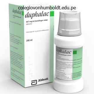
Buy duphalac 100 ml online
Making an "L"formed reduce on the pulley permits a funnelshaped opening to facilitate the repair. By taking one single stitch on the free angle of the flap to reattach the pulley, the milieu interior of the synovial sheath is recreated. Sourmelis described a method using a quantity 8 toddler feeding tube to retrieve the tendon finish by passing the tube across the pulleys and tagging the tendon to the tube. Withdrawing the pulley will convey the tendon by way of the chosen path into the wound for a restore. Suturing Technique Until therapeutic of the tendon occurs, the restore is underneath strain and early mobilization prevents adhesions. The strength of the repair relies upon upon the variety of strands crossing the repair, the dimensions and energy of the material, the knotting property of the fabric and the technique of repair. Many research have targeted on the core stitch and the conclusion is pretty obvious. The more the variety of strands crossing the restore web site, the stronger is the restore and might higher face up to forces placed upon the repair. Locking loops assist to grasp tendon strands and prevents the suture from slipping alongside the longitudinally oriented collagen fibers of the tendon. Grasping the tendon materials within the middle of the reduce finish with a firm bite, in a nice toothed Adson forceps, the suture is utilized to one stump. With apply this can be accomplished without leaving the bite on the tendon tissue. Knots on the surface will attract extra adhesions, and the stiff suture materials will irritate the skin or the synovial lining. Common suture materials used include polypropylene, monofilament nylon and braided artificial polyester. The needle is first handed from the cut end, longitudinally and exited about 8�10 mm on the volarlateral surface of the tendon. A loop is created by passing the needle transversely in a special plane (from the one it exited) exiting from the alternative volarlateral floor. Another loop is created on this aspect such that the two loops, earlier than being pulled securely, appear to be the "ears of mickey mouse" the suture is then passed longitudinally into the tendon. This complete maneuver is greatest accomplished with only one grasp of the tendon substance in the center of the cut surface. The procedure is repeated on the opposite facet and the knot is gently tightened until the tendon ends are approximated. A continuous epitendinous coaptation suture across the circumference of the repaired surfaces, with even, alternating bites approximately 2 mm from the sting, from every stump with 6(0) materials smoothens out the frayed and pouting edges successfully decreasing repair site bulk. The six and eightstrand repairs have been shown to have better power but are difficult to use without particular suture materials on special needles. The looped Ethibond suture is now out there and could additionally be used for six and eightstrand repairs. All efforts are focused towards reaching sufficient restore web site strength at time 0�6 weeks postoperative period. Reattachment of the avulsed profundus tendon in a Jersey finger was traditionally done utilizing pull out sutures via the terminal phalanx tied over a button on the nail of the finger with wonderful outcomes. Suture anchors have simplified these reattachments with out the necessity for pullout sutures. Lately, bioabsorbable micromini anchors are available with robust braided polyblend sutures. Avulsion fractures with large fragments are amenable to screw fixation or could also be stabilized with 26G chrome steel pull out sutures. Those injuries, which have an result on solely 25% thickness of the tendon, could also be gently and easily shaved off. Between 25% and 50% involvement may be handled with solely a operating epitendinous suture.
Duphalac 100 ml low price
Synchronous wrist and digital workout routines may cut back peritendinous fracture adhesions. Early movement of adjoining joints in closed simple metacarpal fractures expedites recovery of motion and strength with out adversely affecting fracture alignment and results in earlier return to work". With rising attention to sports activities at all ranges and with better facilities and propaganda, the number of such accidents in not solely skilled gamers, but additionally the informal gamers is on the rise. The enhance in adventure activities and the hustle and bustle of public transport in crowded metropolitan areas add to the incidence of those injuries. These accidents forestall further participation in sport and are fairly disabling even in activities of daily dwelling. Identifying these accidents early and treating them correctly can result in wonderful recovery and return to the identical level of the game or skilled, recreational and personal exercise. Terminal Interphalangeal Joint Collateral ligament accidents as also dislocations of the terminal interphalangeal joint are uncommon. More widespread injuries related to this joint are avulsion fractures (mallet and jersey finger) or tendon avulsions. Dorsal dislocations are sometimes related to wounds as the skin of the pulp is tethered to the bone to permit agency gripping and stability to hold objects. Dislocations are straightforward to reduce and regain stability, if the adjoining buildings are uninvolved in the harm. Crushing accidents with dislocations are highly unstable and wish extra K-wire stabilization for various intervals. The correct collateral ligament followers out from the neck of the proximal phalanx to insert on the facet of the middle phalanx. The volar plate and the collateral ligaments form three sides of a box which enhances the soundness of the joint. The dorsal capsule is additional strengthened by the central extensor slip and despite this stays essentially the most vulnerable part of the joint. A important lateral impact, usually a glancing harm from a ball, could cause the collateral ligament to get avulsed from the proximal phalanx. Movements are painful and regardless of the passage of time and anti inflammatory medicines, the discomfort persists. Stress X-rays are helpful in diagnosing the harm however the opening up of the joint is very misleading. Through a lateral strategy the collateral ligament is identified and the ligament is freshened. Once the anchor is positioned securely, a Bunnell type suture is run through the ligament finish and the ligament is reattached. Type I (Hyperextension) Avulsion of the volar plate from the bottom of the center phalanx and a minor longitudinal break up in the collateral ligaments is seen. In this, the articular surfaces remain in touch, the center phalanx articulates with the dorsal third of the condyle of the proximal phalanx. Attempts at flexion is usually painful and even passive flexion is at instances not attainable as the ruptured volar plate, a sturdy structure, now edematous, is interposed within the joint. The collateral ligaments are often intact or solely few fibers are concerned in the damage. The insertion of the volar plate, together with a portion of the volar base of the middle phalanx is disrupted. The main portion of the collateral ligaments remains with the volar plate and flexor sheath. In the hyperextension accidents, discount is steady and permits early mobilization. Irreducible or locked hyperextension the volar plate is ruptured and gets interposed between the joint surfaces. With a correct rehabilitation program, full movements could be restored in these circumstances.
Buy generic duphalac
The fracture lines are tough to recognize because of muscular coverage, impaction of the fracture fragments, and hematoma. Among these approaches, generally ilioinguinal is used for anterior column or T-shaped or bicolumnar fractures with delicate comminution within the posterior column, and the Kocher-Langenbeck publicity for posterior column accidents. The incision begins at the posterior superior iliac backbone, proceeds to the greater trochanter, and then continues distally along the femur approximately 10 cm. The fascia lata and the gluteus maximus fascia are divided in line with the incision. The maximus is split along its fibers, with care taken to shield the inferior gluteal nerve. The insertion of the gluteus maximus on the femur could also be divided partially or completely to increase publicity. The gluteus medius and minimus are raised subperiosteally from the ilium and retracted with a Steinmann pin. The superior gluteal vessels and nerve, which emerge from the inner pelvis in this area, should be protected. The fracture fragments usually are discovered attached to the capsule, which varieties the only soft-tissue attachment to the fragments and, therefore, their solely supply of blood supply. To increase the exposure of the roof of the acetabulum, a trochanteric osteotomy could additionally be carried out. Another different is to do a "trochanteric flip" in which the abductor and the vastus lateralis, in continuity, with a small medallion of the trochanter (which is osteotomized in a sagittal plane), are retracted anteriorly to expose the dome of the acetabulum. The benefit of this over a routine trochanteric osteotomy is that the abductors and the vastus lateralis stay in continuity by way of the trochanter, so easy restoration of anatomy is possible. The ilioinguinal approach offers publicity of the whole inner desk of the innominate bone from the symphysis pubis to the anterior side of the sacroiliac joint, including the quadrilateral floor and the pubic rami. Because the abductors muscular tissues remain undisturbed, postoperative rehabilitation turns into quicker. The buildings at risk of injury with this method are the iliac vessels, lymphatic system, femoral nerve, and lateral cutaneous femoral nerve. If a mixed method (anterior and posterior) is deliberate, the floppy lateral position is then preferred. The origin of the stomach muscle tissue from the iliac crest is erased sharply and retracted medially. The iliacus origin is then erased subperiosteally, and the dissection is carried out posteriorly and inferiorly to expose the anterior sacroiliac joint and pelvic brim. Through the medial a half of the incision, the external inguinal ring is identified and the spermatic cord (round ligament in females) is protected with a rubber catheter. The distal flap of the external indirect aponeurosis is raised to reach the mirrored a half of the inguinal ligament. This ligament is incised alongside its size in order to leave 1 mm of the ligament attached to the inner indirect and transversus abdominis origins and the transversalis fascia. The subsequent step is to establish the iliopectineal fascia, which divides the iliopsoas with the femoral nerve (lacuna musculorum) from the exterior iliac vessels (lacuna vasorum). The iliac vessels are retracted, the iliopectineal fascia is recognized after which minimize with blunt-tip scissors from lateral to medial, and the cut is sustained laterally behind the psoas. Next, the psoas with the femoral nerve is retracted as a unit after inserting a rubber catheter round them. After dissecting the iliac vessels with the associated lymphatic tissues as a single unit, along with the areolar tissue around them (to defend the lymphatics and stop postoperative swelling of the limb), these tissues are held together with a 3rd rubber catheter. The periosteum on the internal surface of the pelvis along the quadrilateral plate is now cleared. The exposure is now established by way of three windows, as follows: (1) Retracting the psoas medially allows exposure of the inner iliac fossa, (2) the pelvic brim, and (3) the anterior sacroiliac joint. This exposure is facilitated by flexing and internally rotating the hip to chill out the iliopsoas. The center window is created by retracting the psoas laterally and the vessels medially. This allows the superior pubic ramus and the quadrilateral plate to be visualized. The medial window is seen by retracting the vessels laterally and the spermatic twine medially. This maneuver offers entry to the remainder of the pubic ramus, the pubic symphysis, and the quadrilateral surface.
Order duphalac with paypal
These osteochondral defects, especially when present on the burden bearing areas, may predominantly present as ache, and locking or giving method may be rare. It is essential to notice the next on a plain X-ray: number of loose bodies, their location within the knee joint, the site of origin of that loose body, if attainable. It is important not to confuse the sesamoid bone "fabella" which is commonly current within the lateral head of the gastrocnemius as a free physique. Approach to a Patient There are two important aspects to approaching a patient with free our bodies within the knee: � Excision of the free physique itself, and � Treatment of the positioning of origin of the free body. In these conditions, it might be futile embarking upon surgical removing of those unfastened bodies as the fundamental pathology remains untreated. Proper planning is required as regards the variety of loose bodies and their placement throughout the knee joint. A loose body, which could have been current in the anterior compartment in an old X-ray, might need migrated to the posterior compartment by the time the patient actually decides to bear the surgery. If a brand new film is unavailable, the surgeon might end up spending plenty of time searching for the free physique within the mistaken place. Hence, it could be very important have as latest an X-ray as is possible earlier than embarking upon surgical procedure. It is also helpful to have image intensifier amenities intraoperatively in case a surgeon finds it troublesome to localize a unfastened body at the time of surgery. The surgeon needs to possess both the abilities in addition to the motorized tools (arthroscopic shaver) to have the flexibility to perform the same. If a number of free our bodies are present, then it is sensible to try and take away the smaller ones first through the arthroscopy. Larger portals are likely to leak fluid throughout surgical procedure and therefore must be made as late as is possible in the surgery. The use of commercially out there portal plugs can also assist in decreasing fluid extravasation from these portals. Surgical Treatment Arthroscopy is essentially the most accepted means of tackling a case of unfastened bodies. Not solely does it allow straightforward elimination of unfastened bodies, but it additionally permits a detailed analysis of the anterior in addition to the posterior compartments of the knee joint with minimum morbidity. A variety of arthroscopic graspers must be available for gasping loose bodies of various measurement, consistency and form. Cupped, serrated and low profiles are the varied tips obtainable for grasping round, slippery or skinny flat free bodies respectively. Having a ratchet handle additionally permits the surgeon better freedom in maneuvering the free physique once engaged in the grasper. It is essential to perform a detailed arthroscopic evaluation of the entire joint in every case of free body removing. Occasionally they may be hidden behind synovial folds and therefore it is essential to visualize as well as probe all corners of the knee. Occasionally one might have to resort to making accessory portals in addition to the usual anterolateral and the anteromedial portals. The posteromedial compartment can alternatively be visualized by performing a modified Gillquist maneuver. To prevent damage to the arthroscope, the telescope is replaced by the blunt obturator and gently coaxed into the posteromedial compartment. Once the obturator is replaced by the arthroscope, use of a 70� arthroscope versus a standard 30� arthroscope can also enable a wider area to be examined. A giant variety of studies have been printed indicating advantages, and an equally giant number of research point out in any other case. The truth lies somewhere in between, and affected person selection is an important factor in success of this procedure. The minimally invasive nature of this operation makes it a pure choice for the patient. Overall, it has been proven that arthroscopy has no vital function in osteoarthritis. They must know that arthroscopy, in their case, is primarily diagnostic, and any profit may be a bonus. Sometimes, one does arthroscopy as an investigation previous to deciding whether the patient is appropriate for a unicondylar or total knee substitute.
Order generic duphalac from india
Any rotational malalignment is detrimental to fracture stability, so, if it is present, one must be especially cautious in assessing the steadiness of the reduction and possibly use a 3rd fixation pin. Percutaneous pinning is performed with the maximally flexed and pronated arm resting on the sterile C-arm screen. A easy Kirschner wire is inserted via the lateral condyle, crossing just lateral to the olecranon fossa and interesting the medial humeral cortex. For the lateral view, the arm can be externally rotated on the shoulder whereas flexion and pronation of the elbow is maintained. The medial wire is placed with the arm in 80�90� of flexion; extra elbow flexion may cause the ulnar nerve to subluxate volarward into the trail of the Kirschner wire. Because the lateral wire offers sufficient stability, hyperflexion is now not necessary. The wire is then pushed up the medial column, in order that it crosses the lateral wire proximal to the olecranon fossa. The reduction and wire placement ought to then be checked once more with the C-arm and make sure about bicortical fixation has been carried out. An open discount is indicated in circumstances the place the fracture is irreducible by closed methods or if the brachial artery has been compromised and requires exploration. Lastly, all open supracondylar fractures warrant a surgical debridement of the fracture adopted by stabilization. Wilkins15 reported that buttonholing of the proximal fracture fragment via the brachialis muscle can block discount. In basic, the surgical method ought to be through the area of disrupted periosteum. The neurovascular deficit, if current, must also be considered in planning the surgical strategy. An anterior or anteromedial strategy should be used for a posterolaterally- displaced fracture associated with vascular compromise or a median nerve deficit. In basic, essentially the most versatile strategy is through an anterior transverse incision over the antecubital fossa, with extension of the medial side proximally and the lateral facet distally as needed. The anterior strategy has some nice benefits of permitting direct visualization of the brachial artery and median nerve in addition to the fracture fragments. Once reduction has been achieved, fixation with crossed Kirschner wires is really helpful. Several stories have proven angiography to be an unnecessary take a look at that has no bearing on treatment. All underwent reduction and percutaneous pin fixation with no preoperative angiogram. Blood move to the hand was not restored after the reduction in three of the 17 sufferers, and open exploration was required. In 14 of the 17 patients, blood flow to the hand was restored without issues. The creator concluded that prereduction angiography provides nothing to the management of these injuries. Their indications for arterial exploration have been: � Absence of a palpable pulse after discount with any suggestion of decreased capillary refill, elevated compartment strain or pallor � Total absence on Doppler imaging of a pulse in a nonischemic extremity. Our apply is to admit the kid to the hospital, elevate the limb slightly, and observe her or him for no less than 48 hours. While postoperative protocols range from surgeon to surgeon, a typical regimen calls for a long-arm splint or a cut up long arm solid to control elbow movement and forearm rotation for 4 weeks, followed by pin removal and early vary of motion or continued splinting for additional 1�2 weeks. If a secure closed reduction and an experienced pediatric orthopedic surgeon achieves pinning of the fracture, follow-up might safely be delayed till the day of pin removing. This early follow-up for unstable fractures permits for a repeat closed manipulation and pinning if there has been a loss of reduction. Children who is most likely not reliably examined for compartment syndrome because of younger age or cognitive disability are typically treated emergently, as are kids with full motor and sensory median nerve deficit.
Syndromes
- Do NOT apply a heating pad or hot water bottle to your feet. Avoid hot pavement or hot sandy beaches.
- Problems with the hearing nerve
- Creatinine clearance
- Tetanus
- More lead will leach into hot liquids like coffee, tea, and soups than into cold beverages.
- Your surgeon will make 3 or 4 small cuts, usually no more than 1-inch each, in your belly and side. The surgeon will use tiny probes and a camera to do the surgery.
Generic duphalac 100 ml overnight delivery
The fact that the carpal adjustments are secondary upon the distal radius malposition is easily confirmed by gently dorsiflexing the wrist and getting a lateral view. Correction of the first radial deformity usually corrects the carpal instability and improves function. Chronic modifications within the intercarpal ligaments and the bony surfaces of the carpal bones may immediate the necessity to add procedures to handle these issues concomitantly. The foregoing serves solely to contact upon the huge and ever expanding scope and information regarding carpal instabilities. Differences of opinion amongst the leading staff within the field continue to make it troublesome to perceive as the nomenclatures range as do the concepts. It is crucial that the treating surgeon is conscious of the situation and intervenes early to ensure the best outcomes. A compromise of the wrist actions Carpal InstabIlIty with a discount in the grip strength is inevitable within the extra complex procedures. Vascularity of Lunate the blood provide to the lunate has been studied extensively. There is both a palmar and dorsal blood supply current in 74�100% of the lunate bones. Based on injection studies, extraosseous blood supply to the palmar side of the lunate consists of the radial, ulnar and palmar branches of the anterior interosseous artery combining to type three transverse arches. The direct dorsal blood supply to the lunate arose from the dorsal carpal plexus on the mid-dorsum of the carpus, which is fed by branches from the radial artery and dorsal department of the anterior interosseous artery. Negative Ulnar Variance Ulnar variance is set on a posteroanterior radiograph of the wrist obtained with the forearm in impartial rotation. Early vascular modifications begin with ischemia, subsequent necrosis, and revascularization. Intermediate osseous changes: Lichtman has described this well over the past 33 years. Late chondral adjustments: the modifications identified have been described earlier in the part on classification. The articular cartilage is commonly soft and may be indented, giving the impression that the articular surface has a false floor. It locations on the front of the decision-making course of, the pathoanatomic features of the articular cartilage. In the normal loaded Kienb�ck wrist, when the subchondral bone has collapsed, the body of the lunate is urgent immediately onto the deep surface of the articular cartilage. With normal wrist operate this interface is subjected to appreciable shear forces. Once the lunate articular cartilage has misplaced its assist, secondary adjustments are more likely to happen reasonably quickly. We have witnessed that once the analysis of Kienb�ck disease requires a excessive index of suspicion, particularly in young males presenting with ache and stiffness in the dominant wrist. They may have lowered grip energy and the ache can be isolated to the dorsal lunate. The onset is often insidious, starting with a dull intermittent ache over the central dorsal wrist. The ache could be aggravated by activity and is often relieved by relaxation and immobilization. Some patients could show a radiocarpal effusion signifying underlying synovitis. Staging and Classification In 1977, David Lichtman published his landmark paper on Kienb�ck disease, together with the radiologic osseous classification system. It has been the standard of evaluation to this time, although issues have been raised with regard to its reliability. The grading system assists the surgeon to decide the most effective surgical option, based on the pathoanatomic findings. The use of exterior fixators has been described,23,26 though that is used more generally along side a revascularization procedure27-29 or cancellous bone grafting of the lunate. Reported results have generally been favorable in phrases of relief of symptoms, though the radiographic appearance of the lunate has been usually static. However, issues occurred in 22% of the patients, together with nine patients with delayed union or nonunion.
Buy duphalac 100ml without a prescription
The area of assembly between the secondary pressure trabecular and the compression trabecular within the heart of the head of femur carries the densest portion of cancellous bone, in which an effort is made to interact the tip of the sliding screw. Sometimes, a further cancellous screw fixation above the sliding screw is advised in severely osteoporotic neck and/or basal fractures. Treatment the goal of therapy is to achieve union without deformity and encourage early mobilization to reduce the morbidity and mortality charges and to restore the affected person to his or her preoperative practical status at the earliest attainable time. Comminution the degree of comminution depends on osteoporosis and pressure of damage. When the combined influence of osteoporosis and comminution is considered, the most secure fracture is the two-fragment fractures of porotic bone. Osteoporosis and comorbidities provides to the complexity of the fracture and its administration. Multiple modalities of treatment, implants and surgical techniques should be mastered. The stability after surgical therapy indicates union of fracture with out deformity and good function. Currently, reduction of anteromedial cortex has been given a lot importance to obtain stability. As there are a big number of fracture patterns, comminution and severe osteoporosis make the fracture remedy advanced, subsequently, needs all kinds of implants devices and thoroughly mastering the procedures instrumentation. It is necessary for an orthopedic surgeon to perceive the biomechanics of the assorted implants. Indication for Nonoperative Treatment � An elderly person whose medical situation carries an excessively excessive threat of mortality type anesthesia and surgery � Nonambulatory affected person who has minimal discomfort following fracture � the conservative treatment by skeletal tibial traction may be tried for 8�12 weeks � Intensive medical and nursing care is required to prevent pressure sores, pneumonia, urinary tract infection, thromboembolism, and pin-tract sepsis. Factors underneath the management of surgeon for profitable treatment are: � Good discount � Proper choice of implant � Proper surgical technique, which includes availability of modern operation rooms and whole set of implants and instrumentations � Availability of image intensifier and clear air system (laminar air flow) in operation rooms can be essential. The factors most vital for instability and fixation failure are: � Loss of posteromedial or medial cortical help � Severe comminution � Subtrochanteric extension of the fracture � Reverse indirect fracture � Shattered lateral wall � Bone high quality. There are a lot of articles with meta-analysis and randomized, potential studies; still there are lot of controversies regarding selection of implants and procedures. Screw of sliding within the barrel depends on: � Fracture geometry � Quality of discount � Position of screw in the head and neck of femur � Angle of the barrel and plate � Integrity of lateral wall. The shearing pressure on the femoral head being transferred to the axis of the sliding screw, therefore producing a compressive drive. Note the stress in the facet plate to compress the fracture of the lateral cortex. For this purpose, the top screw is placed higher to acquire distance inside the proximal fragment. Even if the fracture unites, there may be thigh pain, shortening of limb and unhealthy limp Cutout of implant might occur in extreme osteoporotic bone Reverse oblique fracture are unstable fractures. Excessive collapse occurs due to shearing forces and to powerful muscle tissue performing on fragments. Reduction: Patient is positioned in a supine place on a fracture desk, preferably under epidural anesthesia. Reduction of the fragments, especially the anteromedial, is an important prerequisite for good union. Anatomical discount may be achieved by closed methodology by applying traction to over distract. Most fractures are lowered by direct traction, slight abduction and normally 10�15% inside rotation. Occasionally slight exterior rotation may be required for more intensive and comminuted fractures. With picture intensifier the limb should be rotated internally to obtain satisfactory reduction. It is important to check on the lateral projection that the shaft has not sagged posteriorly. Once reduction is achieved, most essential step is to keep it, until definitive fixation is full.
Order 100 ml duphalac otc
Anatomical basis of the danger of radial nerve injury related to the technique of external fixation utilized to the distal humerus. The use of an antibioticimpregnated, osteoconductive, bioabsorbable bone substitute within the remedy of contaminated long bone defects: Early outcomes of a potential trial. Nonunion of the humeral shaft: lengthy lateral butterfly fracture-a nonunion predictive sample Section 19 � � Injuries of Elbow Section Editor: Prakash P Kotwal � � Fractures of Distal Humerus in Adults Sudhir Babhulkar Monteggia Fracture Dislocation Prakash P Kotwal, Mohammed Sadiq Injuries Around Elbow Abhinav Agarwal, Prakash P Kotwal, Mohammed Sadiq Fractures of the Olecranon Prakash P Kotwal, Abhinav Agarwal Dislocations of Elbow and Recurrent Instability Alex Sonaru, Justin Gettings, Guido Marra Fractures of the Radius and Ulna Prakash P Kotwal, Amit Singla Sideswipe Injuries of the Elbow Prakash P Kotwal, Abhinav Agarwal 175 Chapter Fractures of Distal Humerus in Adults Sudhir Babhulkar Introduction Fractures of the distal humerus are relatively uncommon, however are very challenging to manage. The possibilities of functional impairment and deformity are very high following the conservative therapy of such distal intraarticular fractures of humerus and inflexible inner fixation may be difficult to achieve as a outcome of complexity of fracture and osteoporosis. Severe comminution, bone loss and osteopenia predispose the fracture for unsatisfactory outcomes due to insufficient fixation. Over 25% of such fractures develop significant issues in the course of the remedy and few of them may have further surgery. With the arrival of current and modern reconstructive methods, the prognosis of distal humerus fractures has changed. Surgical Anatomy of Distal Humerus the distal humerus consists of the expanded portion of the metaphysis, together with the joint surface for articulation with corresponding surfaces of proximal ulna and radial head. The ulnotrochlear joint moves via a single axis of rotation, flexion extension of elbow joint, whereas radiocapitellar joint is mechanically linked to inferior radioulnar joint attaining forearm rotation of pronation-supination movements. The distal most part of the lateral column is the capitellum, and the distal most part of the medial column is the nonarticular medial epicondyle. The trochlea is the medial most part of the articular phase and is intermediate in place between the medial epicondyle and capitellum. The eminences of the pulley present medial and lateral stability to the straightforward hinge joint. The pulley like trochlea types a central articulating axis and the distal humerus articular phase features architecturally as a tie-arch between the medial and lateral humeral columns-pillars. Restoration of mechanical stability of fracture, distal humerus depends upon recreating this triangle of stability. Pillars and trochlea has the realm of greater bone mass and serves as the primary space for internal fixation. The trochlear axis can be rotated between 3� and 8� with respect to a line connecting the medial and lateral condyles. The articular section just forward from the road of the shaft, lengthy axis of humerus at 40� and features architecturally as the tie arch at the point of most column divergence distally. The medial epicondyle is on the projected axis of the shaft, whereas the lateral epicondyle is projected barely ahead from the axis. They also are likely to underestimate the severity of displacement, degree of articular involvement, and comminution of distal humeral fractures. Traction radiographs could higher delineate the fracture pattern and may be helpful for preoperative planning. In nondisplaced fractures, an anterior or posterior fats pad signal may be present on the lateral radiograph, representing displacement of the adipose layer overlying the joint capsule within the presence of effusion or hemarthrosis. Minimally displaced fractures fracTures of disTal humerus in adulTs might result in a lower within the regular condylar shaft angle of 40� seen on the lateral radiograph. Type A fractures are extraarticular, Type B fractures are partial articular, and Type C are full articular. The classification additionally permits distinction of the extent of the fracture, which is a serious determinant of the complexity of any reconstructive procedure. However, as on today, the controversy nonetheless exists within the administration of intra-articular distal humerus fractures in adults:9 � Whether to operate or treat conservatively A lateral Kocher approach will usually be sufficient to acquire access to the lateral column to enable sufficient fixation. A medial method, posterior triceps reflecting, or transolecranon strategy may be required to provide enough publicity to treat a medial column fracture or a extra comminuted low medial or lateral column fracture. High fractures are more unstable, however easier to reconstruct, because of the bigger fracture fragment, which is able to more easily accommodate internal fixation. Reduction of the fracture is carried out and a formal arthrotomy is usually required to assess the adequacy of the articular reduction. After provisional stabilization of the fracture, multiple interfragmentary lag screws may be adequate to stabilize the fracture in younger individuals and at instances may require buttress plating to help the lag screws.
Cheap duphalac 100ml free shipping
Ligament laxity is common amongst Indian females and Orientals where each the sexes seem to be equally involved. Rotator Cuff Tests Painful arc-A compromise of the rotator cuff function- both complete or partial, because of tear or irritation, results in an inefficient abduction leading to a painful arc. Fallacies � Patients with inside impingement of the rotator cuff, commonly patients with laxity, will experience apprehension during the Crank take a look at but the ache and discomfort shall be felt 2084 Supraspinatus TexTbook of orThopedics and Trauma Empty can test-The arm is placed in 30 degrees of flexion and abduction in the plane of the scapula with the elbow absolutely prolonged and thumb pointing down (Empty can test) toward the floor. The affected person is requested to raise the arm towards resistance utilized by the examiner over the forearm. The empty can position eliminates many of the deltoid motion however patients with weak supraspinatus could recruit the biceps by flexing the elbow. Full can test-The identical test is repeated with the thumb pointing up toward the ceiling. In the presence of a full thickness tear both the empty can and the complete can exams might be positive. In supraspinatus tendonitis, calcific tendonitis or partial tears of the rotator cuff the total can take a look at shall be negative whereas the empty can check may be optimistic. In thin sufferers with wasted deltoid, often one can palpate the defect in the cuff whereas rotating the arm internally and externally. External rotation may also be examined in opposition to gravity by flexing the shoulder and elbow to 90 degrees and inner rotation on the shoulder joint. The patient is then requested to externally rotate towards gravity against resistance. The other potential inside rotators of the humerus (Pectoralis major and Latissimus dorsi) have a limited position in sustaining inside rotation when the arm is positioned behind the back. Also in subscapularis rupture, a rise in the exterior rotation as in comparison with the normal aspect is a contributory discovering. Also a weak subscapularis in abduction suggests a full body tear involving the inferior insertion of subscapularis. Fallacy-Patients with restricted inside rotation as a result of a tight posterior capsule, will naturally expertise ache on stretching during the cross adduction test. Similarly, in suprascapular compression neuropathy, the nerve could be stretched on the cross adduction check leading to pain Paxinos Sign the examiner performs the check for the Paxinos sign with the affected person sitting comfortably on the examining couch and the affected arm by the facet of the chest wall. The examiner then applies strain to the acromion with the thumb, in an anterosuperior path, and inferiorly to the midpart of the clavicular shaft with the index and lengthy fingers. Long Head of Biceps Speed Test the shoulder is ahead flexed in supination with the elbow 30 degrees flexion towards resistance utilized at the forearm. If the nerve is affected at the root degree, more proximally, then the weakness is profound and winging is instantly apparent. The long thoracic nerve can suffer a compression neuropathy in the midaxillary line just proximal to the innervation of the muscle by its numerous branches. The vascular leash of vessels proximally over the course of the nerve from an adherent scar tethering the nerve causing neuropathy of the branches distal to the nerve. Since the branches proximal to the nerve are unaffected the weakness of the muscular tissues is incomplete. Wall Push Test Performing the wall push with both the elbows in full extension will reveal the winging of the medial border of the scapula. In addition, a young level could be elicited on the above described level to reinforce the prognosis. The sample of winging in trapezius weak spot differs from typical serratus anterior weakness. In addition to atypical winging, sufferers have weak point in elevating the scapula and on account of this develop impingement on the shoulder with stiffness. Compression Neuropathy of Suprascapular Nerve Compression neuropathy of the suprascapular nerve is a uncommon and often recognized situation. A lesion within the spinoglenoid notch will invariably affect only the infraspinatus muscle.
Discount duphalac online master card
The head is covered with an impervious sheet which might be caught to the superior neck with micropore tape. After glenohumeral arthroscopy, the arm abduction gadget may be adjusted to place the arm in 20�30� of abduction and impartial flexion in order to facilitate visualization of the subacromial house. No additional equipment required (unlike a head holder and spider within the seashore chair position). Pros and Cons of a Lateral Decubitus Position Pros � Positioning is much less complicated as a lateral position is very commonly utilized in working theaters for hip surgical procedures. Cons � Orientation of the humeral head with the glenoid may be difficult initially. The buttocks and higher trochanter are positioned at the primary break of the working table. The head and back are then elevated to obtain a sitting position with an roughly 20�30� back tilt. The affected person is dropped at the edge of the desk such that the posterior aspect of the shoulder is uncovered till the midscapular space. Head positioners connected to the desk ensure strapping and placement of pads and/or gelfoam such that � the top and neck place are securely maintained through out the process. Alternatively, the head may be placed in a jelly pad and strapped securely with tape. The arm can be � Either positioned free with the elbow resting on a facet assist � Or placed in an arm positioner. This helps management rotation and eliminates the necessity of an assistant doing so if the arm was left free. The anesthetic machine is best positioned at the foot end of the table such that the surgeon and his assistant/s have free access across the shoulder. The neck and head are sealed off with an impervious Udrape such as to keep away from getting wet throughout the procedure. Cons � Posterior and posteroinferior glenohumeral arthroscopy is barely troublesome compared to the lateral place � Requires an accurate desk for the purpose and extra equipment such as the Spyder to place the operating limb. It is located in the gentle spot (the area between the infraspinatus and teres minor) approximately 3 cm inferior and 1 cm medial to the posterolateral acromion. After glenohumeral arthroscopy, the arthroscope can be repositioned via the identical portal into the subacromial area. It can be used as a working portal when the arthroscope is getting used from one of many anterior portals. If one is simply too low (approximately 6�7 cm inferior to the posterolateral acromion), the axillary nerve and circumflex humeral vessels could be at risk. When considered from posterior, this portal enters the joint in the triangle formed by the biceps tendon, humeral head and glenoid, just superior to the subscapularis tendon. It is a crucial working portal whilst performing labral and capsular procedures. It can be used as a viewing portal whilst working within the posterior glenohumeral joint. If one is merely too low (through or under the subscapularis), the subscapularis vessels and cephalic vein are in danger. It is used as a viewing and working portal in rotator cuff and subacromial procedures. If one is merely too distal (approximately 5 cm distal to the lateral acromion) the axillary nerve could be in danger. Basic Portals of Shoulder Arthroscopy A correct understanding of shoulder anatomy is essential to respect correct portal placements. Correctly positioned portals are a key to a easy, dependable and reproducible procedure. Anatomic considerations that may prevent neurovascular harm are: � Anteriorly: Do not stray medial to the coracoid. Portals could be used as: � Viewing portals-can be interchanged during surgical procedure shoulder posiTioning, primary porTals and seaside chair versus laTeral decubiTus posiTion � Rotator cuff restore � Standard posterior portal � Lateral portal � Anterosuperior portal Accessoryportals: Posterolateral Wilmington Neviaser. A comparison of threat between the lateral decubitus and the beachchair position when establishing an anteroinferior shoulder portal: a cadaveric study. Inflatable pillows as axillary assist devices during surgery performed within the lateral decubitus position under epidural anesthesia. Betaadrenergic blockers and vasovagal episodes during shoulder surgery within the sitting position under interscalene block.
Real Experiences: Customer Reviews on Duphalac
Nerusul, 27 years: A fall onto an outstretched hand from a top of only 6 cm is estimated to create an axial joint compression drive at the elbow of 50% of physique weight.
Ali, 42 years: A volar subluxated fracture fragment is probably the one absolute contraindication for dorsal plating (Box 2).
Gunnar, 38 years: A total of 60 limb segments were lengthened-40 tibiae, 12 femura, four radii, one humerus, and three feet.
Deckard, 37 years: However, just a few of the muscles that cross the joint act primarily to transfer the joint, the biceps, brachialis, and brachioradialis flex the elbow.
Amul, 23 years: Flex the arm by 90 degrees at the shoulder and elbow and forcefully internally rotate it to provoke impingement.
Mortis, 21 years: Treatment of posttraumatic radioulnar synostosis with excision and low-dose radiation.
Ashton, 45 years: In no case, should an antegrade femoral nail be inserted to deal with both fractures because of the high danger of displacing the fracture.
Rakus, 49 years: However, the relatively minor episode of trauma and the medical presence of functioning anterior and posterior cruciate ligaments often results in a missed analysis.
10 of 10 - Review by T. Angar
Votes: 41 votes
Total customer reviews: 41
References
- Schoenberg SO, Essig M, Bock M, et al: Comprehensive MR evaluation of renovascular disease in five breath holds, J Magn Reson Imaging 10(3):347-356, 1999.
- Valcarcel D, Sanz MA, Sureda A, et al. Mouth-washings with recombinant human granulocyte-macrophage colony stimulating factor (rhGM-CSF) do not improve grade III-IV oropharyngeal mucositis (OM) in patients with hematological malignancies undergoing stem cell transplantation: results of a randomized double- blind placebo-controlled study. Bone Marrow Transplant. 2002;29:783-787.
- Tomashefski JF, Cramer SF, Abramowsky C, Cohen AM, Horak G. Needle biopsy diagnosis of solitary amyloid nodule of the lung. Acta Cytol 1980;24:224-7.
- Bhavnani SP, Coleman CI, White CM, et al: Association between statin therapy and reductions in atrial fibrillation or flutter and inappropriate shock therapy, Europace 10:854-859, 2008.


