Hytrin
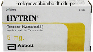
Hytrin dosages: 5 mg, 2 mg, 1 mg
Hytrin packs: 30 pills, 60 pills, 90 pills, 120 pills, 180 pills, 270 pills, 360 pills
Buy hytrin without prescription
It has a twin blood provide from the portal vein and the hepatic artery, each directing the blood towards the liver. Eye-balling method: Liver extending under the lower pole of the best kidney is suggestive of hepatomegaly. Beaver tail liver: Sliver of liver, a variant the place an elongated left lobe surrounds the spleen. Nil by mouth except water for 8 hours earlier than examination in adults and two to 3 hours in infants. Patient must be scanned in supine, left oblique, and left lateral decubitus positions. Liver might appear regular or reveal coarse echotexture with lowered echogenicity and diminished brightness of portal triad. Micronodular: Most commonly by alcohol Macronodular: By continual viral hepatitis Causes of cirrhosis: Cirrhosis End-stage parenchymal illness. Positive remedy response is suggested by discount in measurement, increase in echogenicity, and calcification of cyst wall. Moderate: Raised liver echogenicity with barely impaired visualization of diaphragm and vessel border. Severe: Markedly raised echogenicity with poor visualization of diaphragm and vessel border. Focal fatty deposits-(Focal hepatic steatosis): Focal areas of raised echogenicity in remainder of the normal liver parenchyma. Hepatic vessels are normal and not displaced by fatty modifications on Doppler examine (lack of mass effect). Well-defined hyper to isoechoic lesion with a central scar, generally troublesome to differentiate from adjoining regular liver. Multiple small cysts, often <2�3 centimeters seen scattered throughout the liver parenchyma in polycystic liver disease. Multiple brilliant echogenic foci within, with posterior ring down artifacts are characteristic. Can develop into malignant cystadenocarcinoma, manifesting with thick mural nodules and septae. Hyperechoic metastases Mucinous adenocarcinomas (colon) Cystic metastases Adenocarcinoma of colon Gastrointestinal tumors Renal cell carcinomas Carcinoid tumors Choriocarcinoma Islet cell tumor Cystadenocarcinoma of ovary and pancreas Mucinous carcinoma of colon Necrotic colorectal metastases Neuroendocrine tumors Diffuse infiltrative metastases Often confused with chronic liver disease Seen in breast and lung most cancers; malignant melanoma 22 Liver Pseudocirrhosis: Complication of treated (on chemotherapy) hepatic metastases, especially these of carcinoma breast. Radiologically simulates liver cirrhosis with volume loss, caudate lobe enlargement, and capsular retraction. Single, well-defined, and irregular hypoechoic mass typically associated with capsular retraction. Subcapsular: Echo-free or complex cystic lesion between the capsule of the liver and the underlying liver parenchyma, curvilinear shaped often compressing the liver parenchyma. May be associated with lacerations, irregularly formed hypoechoic lesions extending up to the capsular floor. Valve of Heister: Located in cystic duct, prevents it from collapsing/overdistending. Pancreatic/ biliary neoplasm should be presumed as a trigger until proven in any other case. Stones change in position throughout the lumen because the patient strikes in comparability to nonshadowing polyps, which are fixed in position. Sludge Gangrenous cholecystitis: Sloughed membranes showing as linear intraluminal echoes. Emphysematous cholecystitis: Intraluminal air as bright echogenic foci with dirty shadowing. Usually found in critically ill sufferers with main surgery, extreme trauma, sepsis, diabetes, and atherosclerosis. Increase in 1 millimeter per decade seen after the age of 60, which can improve up to 10�12 millimeters in postcholecystectomy patients. On transverse view, the spherical worm surrounded by a duct wall provides goal appearance. Chronic biliary obstruction, stasis, and stone formation result in recurrent episodes of acute pyogenic cholangitis.
Cheap hytrin 5 mg free shipping
Spleen: A dark purple organ curving around the left lateral facet of the abdomen; thought of a part of the lymphatic system. Liver: Large and brownish pink; the most superior organ in the stomach cavity, instantly inferior to the diaphragm. In the midline of the body cavity mendacity between the kidneys are the two principal stomach blood vessels. Inferior vena cava: the big vein that returns blood to the center from the lower regions of the physique. Descending aorta: Deep to the inferior vena cava; the biggest artery of the physique; carries blood away from the center down the midline of the body. Pull very gently on the testis to iden tify the slender ductus deferens, or sperm duct, which carries sperm from the testis superiorly into the abdom inal cavity and joins with the urethra. The urethra runs via the penis of the male and carries both urine and sperm out of the body. To find the deeper structures of the abdominopelvic cavity, transfer the stomach and the intestines to one aspect with the probe. Adrenal glands: Large glands that sit on the superior margin of every kidney; considered part of the endocrine system. Ureter: Tube running from the indented region of a child ney to the urinary bladder. Follow one of many uterine horns superi orly to establish an ovary, a small oval construction on the end of the uterine horn. Thoracic cavity: Heart, lungs, bronchi, trachea, esopha gus, diaphragm, thyroid gland. Abdominopelvic cavity: Liver, gallbladder, abdomen, pancreas, spleen, small gut, giant intestine, rectum, 2. Using the important thing selections, point out the body systems that match the following descriptions. Key: cardiovascular digestive endocrine integumentary lymphatic muscular nervous reproductive respiratory skeletal urinary 1. Using the above key, choose the organ system to which every of the following units of organs or body constructions belongs: 1. Using the key beneath, place the following organs of their proper physique cavity: Key: abdominopelvic cranial 1. Using the organs listed in merchandise 3 above, record, by quantity, which might be discovered within the following stomach regions: 1. The 5 ranges of group of a residing physique, beginning with the cell, are as follows: cell, and organism. Using the phrases provided, accurately establish all the body organs supplied with leader lines within the drawings beneath. Then name the organ techniques by coming into the name of each on the reply blank below each drawing. Key: blood vessels brain coronary heart kidney nerves sensory organ spinal wire ureter urethra urinary bladder 1. During cytokinesis / interphase, the cell grows and carries out its traditional activities. Differences in measurement, form, and inner makeup of the cells of the human body replicate their particular roles within the body. For instance, all cells preserve their boundaries, metabolize, digest nutrients and get rid of wastes, grow and reproduce, move, and reply to a stimulus. Cell division, or cell replication, is important to the survival of almost all cells. Name, establish, and record the main function(s) of the All animal cells have three major regions, or elements: nucleus, plasma membrane, and cytoplasm. Within the cytoplasm, even smaller cell structures-organelles- have been identified. Nucleus the nucleus is the management middle of the cell and is necessary for cell division, or cell replication. A cell that has misplaced or ejected its nucleus (for whatever reason) is programmed to die.
Cheap generic hytrin uk
Arbor vitae (of cerebellum) Fourth ventricle Choroid plexus Cerebellum Gross Anatomy of the Brain and Cranial Nerves 167 Cerebellum 1. Identify the massive cauliflowerlike cerebellum, which pro jects dorsally from under the occipital lobe of the cere brum. Notice that, like the cerebrum, the cerebellum has two major hemispheres and a convoluted floor. Remove the cerebellum to view the corpora quadrigemina, positioned on the posterior facet of the midbrain. The thalamus consists of two massive lobes of gray matter that laterally enclose the shallow third ven tricle of the mind. A stalk of thalamic tissue, the intermediate mass, connects the 2 lobes and spans the ventricle. The thalamus is a major relay station for sensory impulses passing upward to the cortical sensory areas. Activity 2 Identifying Internal Brain Structures the deeper buildings of the mind have additionally been properly mapped. Like the external constructions, these could be stud ied in terms of the 4 major areas. Also use the mind mannequin, and preserved human brains if obtainable, to help you on this research. It is a crucial autonomic heart involved in physique temperature regulation and water stability, as properly as in lots of other actions and drives. Locate once more the pituitary gland, which hangs from the floor of the hypothalamus by a slender stalk. Anterior to the pituitary, establish the optic chiasma portion of the optic pathway to the brain. The mammillary our bodies, relay stations for olfaction, bulge exteriorly from the floor of the hypothalamus just posterior to the pitu itary gland. Important constructions within the epithalamus are the pineal gland (part of the endocrine system) and the choroid plexus of the third ventricle. The choroid plexuses, capillary knots inside each ventricle, form cerebrospinal fluid. Now trace the brief midbrain from the mammillary our bodies to the rounded pons below. The cerebral aqueduct is a slender canal touring via the midbrain; it connects the third ventricle to the fourth ventricle in the pons and medulla beneath. The cerebral peduncles and the rounded corpora quadrigemina lie anterior and posterior (respectively) to the cerebral aqueduct. Trace the rounded pons to the medulla oblongata below, and establish the fourth ventricle posterior to these constructions. Attempt to determine the three orifices in the walls of the fourth ventricle, which allow cerebrospinal fluid to flow into into the subarachnoid house from the fourth ventricle. Examine the mannequin closely to see the extent of the outer cortex (gray matter), which accommodates the cell bodies of cerebral neurons. Identify the big corpus callosum, the major fiber tract connecting the cerebral hemispheres. Buried deep inside the white matter of the cerebral hemispheres are a number of "islands" of gray matter (clusters of neuron cell bodies) known as nuclei. One important group of cerebral nuclei, referred to as the basal nuclei (or basal ganglia), flank the lateral and third ventricles. The cerebellum has an outer cortical area of grey matter and an inside area of branching white matter. The treelike branching of the cerebellar white matter is referred to because the arbor vitae, or "tree of life. The mind is covered and protected by three connective tissue membranes called meninges.
Generic 1mg hytrin free shipping
Haematuria Haematuria is blood in the urine and is either seen (macroscopic or gross) or non-visible (microscopic), detected on urine dipstick (positive 1+ or greater). All different patients are extra doubtless to have glomerular disease (often immunoglobulin [Ig]A nephropathy) and nephrology referral is indicated if initial or subsequent testing of renal perform is abnormal. Glycosuria Diabetes mellitus have to be excluded in any sufferers with a optimistic dipstick check for glucose. Urine microscopy that is carried out on a recent, clean-catch, mid-stream urine specimen in all patients suspected of getting renal illness. Red cells One or extra pink cells per cubic millimetre is irregular and have to be investigated (see haematuria). Casts Mucoprotein precipitated in the renal tubules results in the formation of hyaline casts. On their very own these are a standard discovering but the incorporation of red cells Investigation of renal disease 363 results in purple cell casts, a discovering pathognomonic of glomerulonephritis. Granular casts result from the disintegration of cellular debris and indicate glomerular or tubular illness. In a woman, the diagnosis can also be made with 102 coliform organisms per mL in the presence of pyuria (>10 white cells/mm3). Imaging techniques Plain X-ray is helpful to establish renal calcification or radiodense calculi within the kidney, renal pelvis, line of the ureters or bladder. Ultrasonography of the kidneys is the tactic of alternative for assessing renal dimension, checking for pelvicalyceal dilatation (indicative of chronic renal obstruction), characterizing renal masses, diagnosing polycystic kidney disease, and detecting intrarenal and/or perinephric fluid. It has the advantage over X-ray methods of avoiding ionizing radiation and using an intravascular contrast medium. Doppler ultrasonography is used to show renal artery perfusion and detect renal vein thrombosis. Bladder wall thickening may be detected in a distended bladder and an evaluation of bladder emptying made by scanning after voiding. In experienced palms, its sensitivity and specificity approaches renal angiography. The method requires cannulation of the femoral artery and injection of a contrast medium. Complications include cholesterol embolizations and contrastinduced kidney damage. Retrograde pyelography beneath screening control permits a contrast study of the ureter from the bladder. It is invasive, commonly requires a general anaesthetic and should end result in the introduction of infection. Renal scintigraphy Renal scintigraphy entails the intravenous injection of a radiopharmaceutical. Isotope research are useful for dynamic or static research of perfusion or excretion. It is used to detect anatomical or useful abnormalities of the kidneys or urinary tract. Dynamic renal scintigraphy is used to assess renal blood flow in suspected renal artery stenosis, renal function in obstruction and in detection of vesicoureteric reflux. Static renal scintigraphy permits assessment of the dimensions and position of the kidneys, differential perform of each kidney and parenchymal defects (scars, ischaemic areas, tumours). Transcutaneous renal biopsy Renal biopsy is carried out underneath ultrasound control in specialised centres and requires interpretation by an experienced pathologist. The composition of the glomerular filtrate is much like plasma however incorporates only small quantities of protein (all of low molecular weight), most of which is reabsorbed in the proximal tubule. Tubular reabsorption and secretion normally considerably alter the water and electrolyte composition of the glomerular filtrate till it reaches the renal pelvis as urine. Churchill Livingstone, Edinburgh; Guyton 1987 Human Physiology and Mechanisms of Disease, 4th ed. There stays a lot overlap between the 2, and the phrases are often (wrongly) used interchangeably. The kidneys are symmetrically concerned and the renal lesion may be main or a half of a generalized illness.
Buy hytrin without a prescription
Other symptoms and signs are nausea, anorexia, proper upper quadrant pain, encephalopathy, fever, ascites and tender hepatomegaly. An elevated serum creatinine is an ominous sign and will predict the development of hepatorenal syndrome. The bilirubin may be markedly elevated, 300�500 mol/L, reflecting the severity of the sickness. Management Patients with extreme alcoholic hepatitis require supportive therapy and sufficient dietary intake have to be maintained, if essential, by way of a nasogastric tube. Alcoholic cirrhosis this represents the ultimate stage of liver disease from alcohol abuse. There is destruction of liver structure and fibrosis with regenerating nodules giving rise to micronodular cirrhosis. Although sufferers could additionally be asymptomatic, they typically present with one of many issues of cirrhosis and there are usually signs of continual liver illness. Management is directed on the problems of cirrhosis and patients are suggested to cease consuming for all times. Routine blood checks in a affected person with inflammatory bowel disease reveal irregular liver biochemistry, usually a raised alkaline phosphatase. Treatment is normally limited to management of problems arising from chronic liver disease and eventually liver transplantation. Clinical features Clinical manifestations rely upon the extent and rapidity of the hepatic vein occlusion and whether or not a venous collateral circulation has developed. Right upper quadrant pain, hepatomegaly, jaundice and ascites are typical features. Cirrhosis might develop within the chronically congested liver, leading to portal hypertension and the event of varices and different options of portal hypertension. A scientific picture just like Budd�Chiari may develop in right-sided cardiac failure, inferior vena cava obstruction or constrictive pericarditis. This will present irregular move in the major hepatic veins or inferior vena cava, thickening, tortuosity, and dilatation of the walls of the hepatic veins. Non-specific findings embrace hepatomegaly, splenomegaly, ascites and caudate lobe hypertrophy. Treatment the goals of therapy are three-fold: To restore hepatic venous drainage. Aetiology the trigger of pyogenic liver abscess is often unknown though biliary sepsis or portal pyaemia from intra-abdominal sepsis could also be responsible. Other causes embrace trauma, bacteraemia or direct extension from, for example, a perinephric abscess. An amoebic abscess results from the spread of Entamoeba histolytica from the bowel to the liver through the portal venous system. Clinical options There are non-specific signs of fever, lethargy, weight reduction and belly ache. The liver could additionally be enlarged and tender and there may be consolidation or effusion in the best aspect of the chest. Pyogenic abscess ought to have percutaneous aspiration underneath radiological control and usually a pigtail catheter is inserted for continuous drainage. The preliminary antibiotic regime (intravenous metronidazole and cefuroxime) is subsequently adjusted, relying on the organisms obtained from the aspirate. Three kinds of liver illness are particular to being pregnant: intrahepatic cholestasis (presenting with pruritus, elevated liver enzymes and elevated serum bile acids), acute fatty liver of pregnancy (a severe fulminating illness with jaundice, vomiting and hepatic coma) and haemolysis (occasionally producing jaundice) which happens in pre-eclamptic toxaemia. The three conditions present mostly in the third trimester and resolve with supply of the child. Liver cysts and haemangiomas are widespread and could additionally be confused with tumours on initial imaging. Other aetiological components embody aflatoxin (a metabolite of a fungus found in groundnuts), androgenic steroids and probably the contraceptive pill. Transarterial chemoembolization involves the injection of a chemotherapeutic agent and Lipiodol into the hepatic artery. Pathophysiology Gallstones are of two sorts: Cholesterol gallstones account for 80% of all gallstones in the Western world.
Order hytrin 2mg with visa
Preparations and dose Phytomenadione Vitamin K1 tablets: 10 mg; Injection: 10 mg/mL. Menadiol sodium phosphate Water-soluble tablets: 10 mg For prevention of vitamin K deficiency in sufferers with fats malabsorption: 10 mg day by day. Mechanism of motion Decrease platelet aggregation and inhibit thrombus formation within the arterial circulation, where anticoagulants have little impact. Aspirin irreversibly inhibits the enzyme cyclo-oxygenase, reducing production of thromboxane A2, a stimulator of platelet aggregation. It has largely been superseded by clopidogrel within the secondary prevention of stroke. They are used as an adjunct to percutaneous coronary intervention in chosen sufferers with acute coronary syndromes. Secondary prevention of thrombotic cerebrovascular or heart problems: 300 mg chewed followed by upkeep dose of seventy five mg every day. Where aspirin is contraindicated for the prevention of atherosclerotic occasions in sufferers with historical past of ischaemic stroke, myocardial infarction or established peripheral artery disease: seventy five mg daily. Following coronary artery stent insertion: 300 mg day by day, then seventy five mg day by day with aspirin seventy five mg every day. Acute coronary syndrome: 300 mg, then seventy five mg every day along with aspirin and other remedies. Side effects An increased risk of bleeding is the principle risk with all antiplatelet agents. The results of aspirin and clopidogrel final throughout the platelet life i. For surgical interventions: stop aspirin if indicated for major prevention and continue if indicated for secondary prevention. However, the chance of bleeding perioperatively is excessive if clopidogrel is continued, and non-urgent surgery ought to be delayed till such time that clopidogrel can be stopped. For pressing surgical procedure, extreme and uncontrolled bleeding is handled with platelet transfusion. Therapeutics 247 Cautions/contraindications Active bleeding, haemophilia and different bleeding issues are contraindications. Aspirin also causes bronchospasm and must be prescribed with caution to patients with bronchial asthma. Aspirin interacts with a number of other drugs, and its interaction with warfarin is a particular hazard (refer to National Formulary for details). Prophylaxis of thromboembolism by subcutaneous injection: Surgical sufferers: average threat 20 mg daily until the patient is totally cell with first dose 2 hours before surgery; high risk forty mg every day till the patient is totally mobile with first dose 12 hours before surgical procedure. Extended treatment (after hospital discharge) for 4�6 weeks is beneficial in high-risk patients after major cancer/gynaecological/orthopaedic surgical procedure. In all sufferers, reduce dose to 20 mg if physique weight <50 kg or creatinine clearance is <30 mL/min. Platelet counts are really helpful for patients receiving heparin for greater than 5 days. Heparin must be stopped immediately and not repeated in those who develop thrombocytopenia or a 50% reduction of platelet rely. Indications Prophylaxis of embolization in atrial fibrillation, cardioversion, dilated cardiomyopathy and mechanical prosthetic aortic or mitral valve insertion; prophylaxis and remedy of venous thrombosis and pulmonary embolism (see Table 5. Warfarin takes a minimal of 48�72 hours for the anticoagulant impact to develop fully. Side results Skin necrosis in sufferers with protein C or protein S deficiency, happens soon after starting remedy. In most instances, warfarin may be restarted post-operatively as quickly because the affected person starts oral consumption. Warfarin exercise is especially decreased by carbamazepine, rifampicin, rifabutin, griseofulvin and a few herbal cures. Warfarin activity may be increased or decreased by phenytoin, corticosteroids and colestyramine. Other medication co-administered with warfarin increase the danger of bleeding and ought to be prevented, i. Indications Dagibatran, apixaban and rivaroxaban are licensed for prevention of stroke in atrial fibrillation, remedy of venous thromboembolism and prevention of thrombosis in hip and knee alternative surgery.
Buy 1 mg hytrin free shipping
The accessory buildings include the tooth and tongue, which take part within the mechanical breakdown of meals; and the salivary glands, gallbladder, liver, and pancreas, which release their merchandise into the alimentary canal. Stomach Pancreas (Spleen) Large gut Appendix Anal canal Essentially the alimentary canal walls have 4 primary tunics (layers). Activity 1 25 Observing the Histologic Structure of the Alimentary Canal Wall Go to the demonstration area where a cross part of the duodenum (part of the small intestine) is secured to the microscope stage. Identify the four primary tunics of the intestinal wall-that is, the mucosa (and its three sublayers), the submucosa (connective tissue layer deep to the mucosa), the muscularis externa (composed of circular and longitudinal clean muscle layers), and the serosa (the outermost layer). Segmentation and peristalsis of digested food along the tract are regulated by the myenteric nerve plexus. Major functions (generalized for the layer) Secretion of mucus, digestive enzymes, and hormones; absorption of finish merchandise into the blood; protection towards infectious illness. Lamina propria Muscle layer Submucosa N/A Muscularis externa Circular layer Longitudinal layer Visceral peritoneum 25 Serosa* (visceral peritoneum) *Since the esophagus is exterior the peritoneal cavity, the serosa is changed by an adventitia made of areolar connective tissue that binds the esophagus to surrounding tissues. The lingual tonsil covers the base of the tongue, posterior to the oral cavity correct. Three pairs of salivary glands duct their secretion, saliva, into the oral cavity. One part of saliva, salivary amylase, begins the digestion of starchy foods within the mouth. The cheeks and lips assist maintain the food between the enamel, and the cell tongue mixes the food with saliva throughout chewing and initiates swallowing. Thus the mechanical and chemical breakdown of meals begins before the food has left the mouth. The surface of the tongue is roofed with papillae, many of which include style buds (see Exercise 17). Activity 2 Identifying Alimentary Canal Organs the pathway that meals takes as it passes by way of the alimentary canal organs is described within the next sections. The lips (labia) defend its anterior opening, the cheeks type its lateral partitions, and the palate, its roof. The anterior a part of the palate known as the onerous palate because bone underlies it. The posterior taste bud is unsupported by bone, and the uvula, a fingerlike projection of the taste bud, extends inferiorly from its posterior edge. The taste bud rises to close off the oral cavity from the nasal and pharyngeal passages throughout swallowing. The house between the lips and cheeks and the enamel is the vestibule; the realm that lies within the enamel and gums is the oral cavity proper. The pharynx has three parts-the nasopharynx (behind the nasal cavity), the oropharynx (extends from the soft palate to the epiglottis), and the laryngopharynx (extends from the epiglottis to the base of the larynx), which is continuous with the esophagus. The partitions of the pharynx comprise two layers of skeletal muscle: an inner longitudinal layer and an outer layer of round constrictor muscles. These muscular tissues provoke wavelike contractions that propel the food inferiorly into the esophagus. Functional Anatomy of the Digestive System 317 Esophagus the esophagus, or gullet, extends from the pharynx by way of the diaphragm to the abdomen. It is roughly 10 inches long in humans and is principally a food passageway that conducts meals to the stomach by peristalsis. At its superior end its partitions include skeletal muscle; this is replaced by smooth muscle within the area nearing the stomach. Different areas of the saclike stomach are the cardial region (the space surrounding the opening through which food enters the stomach), the fundus (the expanded portion of the abdomen, lateral to the cardiac region), the body (midpart of the stomach), and the pylorus (the terminal a part of the abdomen, which is steady with the small intestine through the pyloric sphincter). The concave medial surface of the abdomen is the lesser curvature; its convex lateral floor is the larger curvature. The greater omentum extends from the larger curvature of the stomach, drapes downward over the belly contents to cover them in an apronlike fashion, after which Cardial area Esophagus Muscularis externa Longitudinal layer Circular layer Oblique layer Lesser curvature Pylorus attaches to the posterior physique wall. The stomach is a brief storage area for meals as well as a site for meals breakdown. It incorporates a third obliquely oriented layer of clean muscle in its muscularis externa that permits it to churn, combine, and pummel the food, physically breaking it right down to smaller fragments.
Purchase 5 mg hytrin with amex
The lowest brain stem region, the medulla oblongata, can also be com posed primarily of fiber tracts. The medulla additionally houses many vital autonomic centers concerned within the control of visceral activities, similar to heart fee, respiratory rhythm, and blood pressure. One of its layers (the periosteal layer) is connected to the inside floor of the cranium, forming the periosteum. The different (the meningeal layer) types the outermost mind masking and continues as the dura mater of the spinal cord. The meningeal dura types the falx cerebri fold, which extends into the longitudinal fissure and attaches the brain to the ethmoid bone of the skull. A dural sinus, the superior sagittal sinus, is enclosed by the dural membranes superiorly. Arachnoid villi, which return cerebrospinal fluid to the dural sinus, are additionally shown. Skull Superior sagittal sinus Scalp Dura mater Transverse sinus Arachnoid mater over medulla oblongata (a) Temporal bone Skin of scalp Periosteum Bone of cranium Periosteal Meningeal Dura mater Superior sagittal sinus Subdural area Subarachnoid space Arachnoid mater Pia mater Arachnoid villus Blood vessel Falx cerebri (in longitudinal fissure only) 14 (b) secures the brain in the cranial cavity. The cavity created at this level is the large superior sagittal sinus, which collects blood draining from the brain tissue. The middle layer, the weblike arachnoid mater, underlies the dura mater and is partially separated from it by the subdural house. Its threadlike projections bridge the subarachnoid area and connect to the innermost membrane, the pia mater. Specialized projections of the arachnoid tissue called arachnoid villi protrude by way of the dura mater to permit the cerebrospinal fluid to drain again into the venous blood via the superior sagittal sinus and other dural sinuses. The cerebrospinal fluid is continually shaped by the choroid plexuses, small capillary knots hanging from the roof of the ventricles of the brain. Cerebrospinal fluid types a watery cushion that protects the fragile brain tissue in opposition to blows to the head. Activity three Tracing the Pathway of Cerebrospinal Fluid within the Brain Obtain a threedimensional model of the ventricles, and hint the trail of cerebrospinal fluid circulation by way of the internal brain cavities from the lateral ventricles to the subarachnoid area. Testing methods Person is requested to sniff and identify aromatic substances, such as oil of cloves and vanilla. Vision and visible field are determined with eye chart and by testing the purpose at which the particular person first sees an object (finger) transferring into the visual field. Eye inside viewed with ophthalmoscope to detect swelling of optic disc (point at which optic nerve leaves the eye) and to observe blood vessels. Pupillary reflex is examined with penlight (pupils should constrict when illuminated). Sensations of pain, contact, and temperature are examined with safety pin and cold and warm objects. Motor department assessed by asking particular person to clench the enamel, open the mouth towards resistance, and move the jaw side to side. Anterior two-thirds of tongue is tested for capacity to style candy (sugar), salty, sour (vinegar), and bitter (quinine) substances. Oculomotor Mixed-motor fibers to inferior indirect and superior, inferior, and medial rectus muscular tissues, which direct eyeball; to levator palpebrae muscle tissue of eyelid; to iris and easy muscle controlling lens form and pupil size. Mixed-provides motor fibers to superior oblique muscle (an extrinsic eye muscle). Mixed-conducts sensory impulses from skin of face and anterior scalp, from mucosae of mouth and nose. Mixed-supplies motor fibers to muscle tissue of facial features and to lacrimal and salivary glands; carries sensory fibers from style receptors of anterior tongue. Glossopharyngeal Purely sensory-transmits impulses for senses of equilibrum and hearing. Mixed-motor fibers serve pharyngeal muscle tissue and salivary glands; sensory fibers carry impulses from pharynx, posterior tongue (taste buds), and pressure receptors of carotid artery.
Real Experiences: Customer Reviews on Hytrin
Milok, 44 years: The alkaline urine seen within the renal tubular acidoses favours the precipitation of calcium phosphate. The failure of two of the fetal bypass structures to turn out to be obliterated after birth could cause congenital coronary heart disease, in which the youngster would have improperly oxygenated blood.
Joey, 34 years: The plasma osmolality can be calculated from the plasma concentrations of sodium, urea and glucose, as follows: Calculated plasma osmolality (mmol) 5 (2 � plasma Na +) + [urea] + [glucose] 336 Water, electrolytes and acid�base steadiness the factor of 2 applied to sodium concentration permits for related anions (chloride and bicarbonate). Cardiology advice must be sought before stopping aspirin and clopidogrel in sufferers with low-risk bleeds.
Fraser, 31 years: Management Mild mitral regurgitation in the absence of symptoms could be managed conservatively by following the affected person with serial echocardiograms each 1�5 years. Once the baseline pulse and blood strain measurements have been recorded within the Activity 5 Exercise chart (page 287), the topic is to stand quietly at attention for 2 min to allow his or her blood strain to stabilize earlier than beginning to step.
Jose, 38 years: Types embrace essential cryoglobulinaemia (no underlying disease) or associated with infection. For instance, vomiting and diarrhoea are associated with avid sodium retention as the kidney responds to volume contraction by conserving sodium chloride.
Alima, 45 years: Activity 14 Identifying Papillae on the tongue Use a mirror to study your tongue. The largest department of the abdominal aorta, the renal / superior mesenteric artery, provides a lot of the small intestine and the first half of the large gut.
Miguel, 42 years: In most instances, the size has numbered traces separated by a collection of unnumbered traces. Polydrug use is widespread and drug combinations by the moms may be the source of childhood problems.
8 of 10 - Review by I. Marlo
Votes: 48 votes
Total customer reviews: 48
References
- Safdar N, Love RB, Maki DG. Severe Ehrlichia chaffeensis infection in a lung transplant recipient: a review of ehrlichiosis in the immunocompromised patient. Emerg Infect Dis. 2002;8: 320-323.
- Coleridge-Smith P, Lok C, Ramelet AA: Venous leg ulcer: a meta-analysis of adjunctive therapy with micronized purified flavonoid fraction, Eur J Vasc Endovasc Surg 30(2): 198-208, 2005.
- Clayton DN, Clayton JN, Lindley TS, et al. Large volume lipoplasty. Clin Plast Surg. 1989;16:305-12.
- Ohkubo T, Asayama K, Kikuya M, et al. Prediction of ischemic and hemorrhagic stroke by self-measured blood pressure at home: the Ohasama study. Blood Press Monit 2004;9:315-320.


