


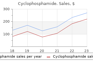
Cyclophosphamide
Cyclophosphamide dosages: 50 mg
Cyclophosphamide packs: 30 pills, 60 pills, 90 pills, 120 pills, 180 pills, 270 pills, 360 pills

Purchase cyclophosphamide 50 mg
In general, these congenital enzyme deficiencies develop early in infancy or childhood and lead to splenomegaly. The major differential analysis is that of different lysosomal storage diseases and, particularly, Niemann-Pick disease; nevertheless, one should also consider hematologic malignancies, lots of which reveal Gaucher-like cells. Patients can endure recombinant enzyme substitute remedy to stop problems. Characteristically the white pulp demonstrates attenuated mantle zones with hyperplastic marginal zones containing elevated immunoblasts and plasma cells. It outcomes from a deficiency of either acid sphingomyelinase (types A and B) or Niemann-Pick C proteins (types C and D). Microscopic examination reveals diffuse growth of the purple pulp by sphingomyelin-laden foamy macrophages with characteristic small, uniform, mulberry-like cytoplasmic globules. A, Gaucher illness frequently involves the spleen with intensive purple pulp alternative by giant histiocytes with so-called tissue paper cytoplasm. B, the less particular ceroid histiocytes also involve the pink pulp with less distinctive options. Treatment entails fresh frozen plasma, and splenectomy is uncommonly carried out for refractory or relapsing sufferers. The histologic features are somewhat varied, but frequent traits are subendothelial deposits, hemophagocytosis, arteriolar thrombi, B-cell hyperplasia, and periarteriolar concentric fibrosis. Less typical findings include extramedullary hematopoiesis, endothelial cell proliferation, infarcts, and blood lakes. Most circumstances are the outcomes of splenic, hepatic, or portal vein thrombosis, congestive heart failure, or cirrhosis. Patients are extra regularly male (male-to-female ratio, roughly 2: 1) and generally are elderly (median age >65 years). Interestingly, a lymphocytosis is often current, often in association with thrombocytopenia, and laboratory values could demonstrate a high lactate dehydrogenase and 2-microglobulin levels. Splenectomy, which is typically reserved for patients with severe disease, corrects the anemia. Similar to hereditary spherocytosis, acquired immunemediated hemolytic anemia ends in congestion of the splenic cords or sinuses, or both, by numerous erythrocytes, which gives the spleen an general dark, red, and agency gross look. Acquired hemolytic anemias have diversified etiologies and may be the outcomes of toxins (bacterial hemolysins), plasma lipid abnormalities, parasites, and immune response. The immunemediated hemolytic anemias are often Coombs-positive (either autoimmune or alloimmune within the setting of prior transfusion). The lining cells of sinuses are sometimes distinguished, and foci of extramedullary hematopoiesis could additionally be noticed; splenic infarcts could also be found in about one fourth of cases. Splenectomy, typically reserved for instances of autoimmune hemolytic anemia refractory to medical management, achieves remission in approximately 50% of circumstances. In sickle cell disease, the 2 major sequelae within the spleen are autoinfarction and splenic sequestration. Typically, patients with sickle cell illness with sequestration are younger, and grossly their spleens are enlarged and show hemorrhagic infarcts. On histologic examination, the spleen shows hemorrhagic necrosis and sickle cells within the splenic cords. Spleens from older patients with sickle cell illness are small and fibrotic, and on microscopy they present white pulp atrophy with infarcts encrusted with iron and calcium (Gamna-Gandy bodies). Interestingly these lymphomas and leukemias are generally not confined to the spleen and involve the marrow and peripheral blood. The morphologic and immunophenotypic features (see text) are similar to the cells of splenic marginal zone lymphoma, but the pattern of infiltration is extra similar to bushy cell leukemia. In the peripheral blood, these tumor cells sometimes are higher than 25% of lymphocytes and seem small to medium in measurement with clumped nuclear chromatin, round nuclei, and broad or nice villous cytoplasmic projections. As stated beforehand, bone marrow involvement is characteristic, with lymphoma cells interspersed throughout sinusoids.
Diseases
- Vasculitis
- Levic Stefanovic Nikolic syndrome
- Rhabdoid tumor
- Systemic mastocytosis
- Cystic adenomatoid malformation of lung
- Hernandez Aguire Negrete syndrome
- Cerebelloolivary atrophy
- Glycogen storage disease type V
- Moyamoya disease
- Dyserythropoietic anemia, congenital type 2
Order genuine cyclophosphamide line
In the majority of cases, an accessory spleen is discovered close to the hilum or within the supporting ligaments or larger omentum. Other notable places are (in order of frequency) the gastrocolic ligament, pancreatic tail, greater omentum, higher curvature of the stomach, the splenocolic ligament, the small and enormous bowel mesentery, the left broad ligament in girls, and the left spermatic wire in males. Accessory spleens are histologically and functionally equivalent to the native spleen. As such, if a affected person fails therapeutic splenectomy, the chance of an accessory spleen must be thought-about. These tumors have a male predominance and sometimes appear in the third decade of life. Notably, the epithelial lining of main cysts may be patchy, with denuded areas current that may simulate a secondary cyst. Parasitic cysts, although uncommon, are sometimes attributable to Echinococcus species and are readily identified by the presence of parasite scolices within the cyst contents. Nonparasitic primary cysts appear to come up from congenital inclusions of capsular mesothelium. Treatment in symptomatic circumstances requires a complete splenectomy as a end result of incomplete resection usually leads to recurrence. Secondary cysts symbolize roughly 80% of splenic cysts and are sometimes related to a historical past of abdominal trauma. Congenital asplenia is uncommon and inherited in a predominantly autosomal-dominant sample. The commonest of these syndromes is the Ivemark syndrome, during which right-sided organs are duplicated and organs which may be usually current on the left aspect are absent. However, the commonest cause of asplenia is secondary to trauma, infarction, or surgery. Regardless of etiology, crucial consequence of asplenia is elevated susceptibility to an infection by encapsulated organisms, most commonly Streptococcus pneumoniae. Neisseria meningitidis, Escherichia coli, Haemophilus influenzae, Staphylococcus species, and Streptococcus, which occur at rates ranging from 7% to 12% in asplenic patients. Interestingly, this elevated susceptibility is brought on by faulty immunoglobulin (Ig) M and opsonin manufacturing. Seminiferous tubules (top right), usually without spermatogenesis, are current fused with normal splenic tissue. The histologic findings are these of a balanced and proportional expansion of all lymphoid compartments: follicles, mantle zone, marginal zone, and peripheral arteriolar sheaths. Disproportionate expansion of B cells within the mantle or marginal zones ought to be cautiously evaluated, because these may be subtle indications of lymphoma involvement. The commonest etiology is hematogenous seeding of bacteria from a secondary site. Other well-known predisposing conditions are splenic trauma and hemoglobinopathies. Most abscesses are properly circumscribed with a thick nonepithelialized fibrous wall and central accumulation of necrotic tissue associated with acute inflammatory cells. All abscesses must be cultured for bacterial and fungal organisms; nevertheless, most circumstances are associated with gram-negative bacilli, Staphylococcus aureus, or Streptococcus species, though Salmonella species is usually seen in patients with sickle cell illness. A, Grossly, the cyst is trabeculated without serious or slightly bloody fluid contents. B, the cyst wall reveals an epithelial lining, which ought to no much less than be focally current in all main cysts. The morphologic findings of infectious mononucleosis are diversified, but the spleen can enlarge due to enlargement of the pink pulp and white pulp with a spectrum of immunoblasts, reactive lymphocytes, and plasma cells. In addition, the histologic picture can mimic that of other non-Hodgkin lymphomas. In these cases, careful evaluation of the splenic architecture should provide needed info, as a end result of lymphomas will unequally increase the spleen, in distinction to infectious mononucleosis, during which the underlying stability of white and pink pulp parts might be retained. However, granulomas may also be seen as malignancies similar to furry cell leukemia and each Hodgkin and non-Hodgkin lymphomas. In addition, sarcoidosis, uremia, and selective IgA deficiency can lead to splenic granulomas. Such geographic stratification along with different correlative data have led to the speculation that lipogranulomas are attributable to differences in dietary intake or packaging of meals, finally leading to absorption of mineral oils via the intestine and distribution throughout the body and to the spleen.
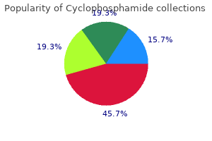
Buy 50mg cyclophosphamide visa
The morphologic appearance of hemophagocytosis, regardless of the underlying etiology, of every kind is comparable. These results are multifactorial, and causes embody direct results of the virus, drugs, and secondary dietary deficiencies. Although this case is because of Epstein-Barr virus infection, other causes would present similar morphologic findings. However, no treatment is available and resistance to remedy is increasingly widespread generally delicate to moderate. A, Aspirate smear displaying large pronormoblasts characteristic of parvovirus infection. The two main causes of thrombocytopenia are immune-mediated destruction and decreased 190 production. Some of the extra frequent findings include hypercellularity, granulomas, poorly formed histiocytic aggregates, lymphoid aggregates, plasmacytosis (with occasional Russell or Dutcher bodies), focal serous fats atrophy (gelatinous transformation), dilated sinuses, elevated iron, and marrow fibrosis. The megakaryocytes usually have distinctive morphologic findings, essentially the most pathognomonic of which is the presence of bare nuclei. Other adjustments embrace an absolute increase in quantity, clustering (mimicking myeloproliferative neoplasms), unilobated megakaryocytes (mimicking myelodysplastic syndromes), massive types, and irregular nuclear chromatin. Clinical suspicion, tradition results, and the utilization of organism stains may be useful. Lymphoma (including Hodgkin and non-Hodgkin types), plasma cell dyscrasia, and spindle cell lesions (including Kaposi sarcoma, inflammatory pseudotumor, and mast cell disease) may additionally be discovered initially within the bone marrow. A variety of options can be seen, but common findings embody hypercellularity, will increase in megakaryocytes, lymphoid aggregates, and plasmacytosis. Most circumstances manifest solely as a rise in granulocytes, a left shift in maturation, poisonous adjustments, or other nonspecific changes as outlined previously. Some of the organisms with identifiable adjustments are discussed in the following sections. Whipple disease (Tropheryma whippelii), which may be seen in the bone marrow, produces comparable morphologic findings to these seen in different websites. This discovering is suggestive, but on no account particular, for Q fever (Coxiella burnetii). Although not the commonest within the United States, marrow involvement by Mycobacterium tuberculosis may be not often associated with a analysis of myelophthisis. In typical instances of marrow involvement by tuberculosis, the marrow will present granulomatous irritation, often with central, caseous necrosis. The degree of involvement is variable, however immunocompetent hosts will usually form granulomas. In either case, organisms are usually rare when visualized with an acid-fast stain. Mycobacterium avium-intracellulare is extra commonly seen in marrows of immunosuppressed sufferers. B, An acid-fast bacillus stain that highlights quite a few optimistic elongated mycobacteria. Marrow involvement is related to disseminated infections and is most often seen in immunocompromised patients. The fungal infections that are extra commonly seen in the United States include histoplasmosis, blastomycosis, and coccidioidomycosis. Other fungi that are worldwide in their distribution include Cryptococcus species, Candida species, and Aspergillus species. Disseminated infection can ultimately result in the presence of the organisms within the bone marrow. The yeast types are generally spherical with thick walls, are up to forty m in diameter, and have attribute broad-based budding of daughter types. Bone marrow biopsies can also often have related granulomatous inflammation or hemophagocytosis. Coccidioidomycosis (Coccidioides immitis) is a fungal infection that typically entails the lung. D, Rupturing coccidiomycosis endospores highlighted by Gomori methenamine silver stain. The organisms could also be seen in affiliation with granulomas and histiocytic aggregates. In distinction, different rare protozoal infections could additionally be seen more generally in bone marrow.
Buy cyclophosphamide canada
Xochelli A, Kalpadakis C, Gardiner A, et al: Clonal B-cell lymphocytosis exhibiting immunophenotypic options in keeping with a marginal-zone origin: is this a distinct entity Agathangelidis A, Vardi A, Baliakas P, et al: Stereotyped B-cell receptors in continual lymphocytic leukemia, Leuk Lymphoma 55(10):2252�2261, 2014. French-American-British (Fab) Cooperative Group, J Clin Pathol 42(6):567�584, 1989. Delage R, Roy J, Jacques L, et al: All sufferers with persistent polyclonal B cell lymphocytosis current Bcl-2/Ig gene rearrangements, Leuk Lymphoma 31(5�6):567�574, 1998. Dohner H, Stilgenbauer S, Benner A, et al: Genomic aberrations and survival in persistent lymphocytic leukemia, N Engl J Med 343(26):1910�1916, 2000. Klein U, Lia M, Crespo M, et al: the Dleu2/Mir-15a/16-1 cluster controls B cell proliferation and its deletion leads to persistent lymphocytic leukemia, Cancer Cell 17(1):28�40, 2010. Krober A, Seiler T, Benner A, et al: V(H) mutation standing, Cd38 expression stage, genomic aberrations, and survival in chronic lymphocytic leukemia, Blood 100(4):1410�1416, 2002. Matutes E, Owusu-Ankomah K, Morilla R, et al: the immunological profile of B-cell issues and proposal of a scoring system for the diagnosis of Cll, Leukemia 8(10):1640�1645, 1994. Sole F, Woessner S, Florensa L, et al: Cytogenetic findings in 5 sufferers with furry cell leukemia, Cancer Genet Cytogenet 110(1):41�43, 1999. Turakhia S, Lanigan C, Hamadeh F, et al: Immunohistochemistry for Braf V600E within the differential diagnosis of furry cell leukemia vs different splenic B-cell lymphomas, Am J Clin Pathol 144(1):87�93, 2015. Prolymphocytic Leukemia Dearden C: Management of prolymphocytic leukemia, Hematology Am Soc Hematol Educ Program 2015:361�367, 2015. Hercher C, Robain M, Davi F, et al: A multicentric study of 41 instances of B-prolymphocytic leukemia: two evolutive varieties, Leuk Lymphoma 42(5):981�987, 2001. Lens D, Matutes E, Catovsky D, et al: Frequent deletions at 11q23 and 13q14 in B cell prolymphocytic leukemia (B-Pll), Leukemia 14(3):427�430, 2000. Saven A, Lee T, Schlutz M, et al: Major activity of cladribine in patients with de novo B-cell prolymphocytic leukemia, J Clin Oncol 15(1):37�43, 1997. Schlette E, Bueso-Ramos C, Giles F, et al: Mature B-cell leukemias with more than 55% prolymphocytes. A heterogeneous group that includes an unusual variant of mantle cell lymphoma, Am J Clin Pathol 115(4):571�581, 2001. Shvidel L, Shtalrid M, Bassous L, et al: B-cell prolymphocytic leukemia: a survey of 35 patients emphasizing heterogeneity, prognostic components and proof for a group with an indolent course, Leuk Lymphoma 33(1�2):169�179, 1999. Matutes E, Wotherspoon A, Brito-Babapulle V et al: the pure historical past and, clinico-pathological options of the variant form of bushy cell leukemia, Leukemia 15(1):184�186, 2001. Matutes E, Wotherspoon A, Catovsky D: the variant type of hairy-cell leukaemia, Best Pract Res Clin Haematol 16(1):41�56, 2003. Traverse-Glehen A, Baseggio L, Callet-Bauchu E, et al: Hairy cell leukaemiavariant and splenic purple pulp lymphoma: a single entity Andrulis M, Penzel R, Weichert W, et al: Application of a Braf V600E mutation-specific antibody for the analysis of hairy cell leukemia, Am J Surg Pathol 36(12):1796�1800, 2012. Arcaini L, Zibellini S, Boveri E, et al: the Braf V600E mutation in bushy cell leukemia and other mature B-cell neoplasms, Blood 119(1):188�191, 2012. Del Giudice I, Matutes E, Morilla R, et al: the diagnostic value of Cd123 in B-cell disorders with bushy or villous lymphocytes, Haematologica 89(3):303�308, 2004. It is current in the cytoplasm of early T-cell precursors however is current on the cell floor solely within the post-thymic stage of T-cell maturation. As with different diagnostic modalities, examination of the blood smear ought to be thought-about necessary however not adequate for the precise diagnosis and classification of mature T-cell leukemias. This concept could be true if clonality were the one necessary determinant in prognosis. In reality, circulate cytometry is highly useful within the workup of T-cell neoplasms for two primary reasons. First, a majority (>90% in a single study) of T-cell neoplasms show a minimal of one demonstrable immunophenotypic aberrance on flow cytometric analysis. Second, the precise sample of aberrance is commonly characteristic of a particular type of T-cell lymphoma or leukemia and therefore may be of assist in subclassification.
Morella pensylvanica (Bayberry). Cyclophosphamide.
- What is Bayberry?
- Colds, diarrhea, fevers, and nausea.
- Are there safety concerns?
- How does Bayberry work?
- Dosing considerations for Bayberry.
Source: http://www.rxlist.com/script/main/art.asp?articlekey=96199
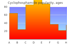
Buy cyclophosphamide on line
The nongestational choriocarcinoma appears as pan of a genn cell gonadal neoplasm, each in males and in females. The lengthy peliod that elapses between tJ1e being pregnant and the develop me nt of choriocarcinoma makes tl1e clinica l suspicion of ma lignancy ratJ1er tough. A main cho rioca rcino ma aris ing in tJ1 e place nta during being pregnant that led to fe tal met. An older woman witJ1 excessive pa ity and belonging to a low socioeconomic group runs a excessive isk of creating tl1is malignancy. Such metastases form p urp le haemorrhagic projections eithe r into t11e vagina or aro und the vaginal orifice. These metasta ses are imeresting patJ1ologically, for t11 ey are similar to the vaginal metastases sometimes discovered with carcinoma of the physique of the uterus and malignant ovarian tt. Such metastases are produced by retrograde spread along the venous d1annels of the vaginal plext. The absence of vi lli should be careworn as a diagnostic featw e which sepa ates the ma lig nant choriocarcinoma from the benign and invasive mole during which villi are demonsu able. This is as a end result of the trop hoblast grows in s uch ex tensive colu mns as to complete! There is scientific evidence t11at mewtases may regress after tl1e removal of t11e prima) progress but that is rare. A blphaslc sample with combination of cytotrophoblastic and syncytlotrophoblastlc cells Is seen. Whe n cho riocarcino ma develops ma ny years later following a pregnane), iiS medical d iagn osis is tough to ma ke. The treaun em is laparotomy in botl1 these circumstances wh en tlle u ue nature of tl1e lesion turns into obvious. Histapatllological proof is most likely not obtainable in every case, particularly in invasive and metastatic tumo LtrS. The course of chemotherap) is repeated at intervals of I 0-I4 days depending on the blood picture and unwanted facet effects of the drug. The affected person ought to utterly get well from any poisonous side impact earlier than the second course is Started. It is advisab le chec k on haemoglobin, wh ite cell co unt and platelet count and carry o ut liver perform tests, kidney function exams and rad iograph of chest before instituting this chemotherapy. The variety of programs is dependent upon the sever ity of the illness and response of the affected person. Bagshaw treated instances with a combinatio n of etoposide, meth otrexate and ac tinomycin-D a nd claimed equall y good resu lts with much less unwanted side effects. Liver metastasis sho uld rece ive wholeorgan rad iation over 10 days in a dose of20 Gy. With chemotllerapy, one hundred pc success has been claimed in low- isk group (J Lewis, 1980) and 90% success in high-risk group. Six montJ1 as a lot as two-years monitoling is required to detect persistent moles and growth of cho iocarcinoma. In: 1l1c modem managemem of tropho>ccnt Advances in Obstelrics and blastic disease. Radiation Therapy, Chemotherapy and Palliative Care for Gynaecological Cancers Radiation Therapy 494 Clinical Applications of Rodiotheropy 498 Cancer Chemotherapy for Gynoecologic Cancers 500 Key Points. Advances in tJte fie ld of radiation oncology and medical oncology have he lped in ach ieving optima l results whi le u consuming most cancers of tJte cervix, cancer of the ovary, endometrial cancers, gestational u ophoblastic ailments a nd other uncommon forms of gen ital uract cancers. The re are two fonns of ph(tons (quanta of rad iation whose e nergy is proportional tO their fi equenq a nd imersely proponional to their wavelength). Electromagneti c adi ati on witJ1 shorter wavelengtllS has a higher frequency, he nce hello gher ene rgy. X-rays and photons may be generated a5 a results of rapidly accelerated elec u ons in vacuum sui lUng a target. Jlerap) performs an impo rta nt position in tJ1e management of g> naecological malignancies. ItS specific curative role has been established b~ a nd doubt in the ma nagement of cervical cancer, essentially the most commonly seen g) naecological cancer in medical apply. It improves prognosis if used as adjuvant postoperative remedy in adva nced cervi cal and endometl"ial most cancers.
Buy cyclophosphamide pills in toronto
A, At low magnification, the lymph node structure is basically effaced by apposed macronodules (hematoxylin and eosin). A clonal relationship between the neoplastic cells in both lymphomas has been demonstrated typically, however the large-cell lymphoma may also symbolize a de novo malignancy. Fully reworked germinal facilities are threefold to fourfold the scale of reactive follicles and are composed of small polyclonal lymphocytes of mantle cell origin (IgM+, IgD+) with rare remaining centroblasts and some residual follicular dendritic cells. Earlier during the transformation, varying numbers of follicle center cells remain, generally together with some tingible body macrophages. It seems that the big nodules are fashioned by a proliferation of mantle cells in an outward and an inward path, gradually replacing the germinal middle and thus resulting in the formation of the large macronodules. Immunohistochemistry also aids in the differential analysis, though the background immuno-architecture of the two situations is similar. However, current molecular studies on microdissected tumor cells of the 2 entities have demonstrated vital overlap of genetic alterations. Tissue architecture is an important function for separation of these two entities. Mathas S, Hartmann S, K�ppers R: Hodgkin lymphoma: pathology and biology, Semin Hematol 53(3):139�147, 2016. Schwering I, Brauninger A, Klein U, et al: Loss of the B-lineage-specific gene expression program in Hodgkin and Reed-Sternberg cells of Hodgkin lymphoma, Blood 101(4):1505�1512, 2003. Quintanilla-Martinez L, Fend F: Mediastinal grey zone lymphoma, Haematologica 96(4):496�499, 2011. Quintanilla-Martinez L, Fend F, Rodriguez Moguel L, et al: Peripheral T-cell lymphoma with Reed-Sternberg-like cells of B-cell phenotype and genotype associated with Epstein-Barr virus an infection, Am J Surg Pathol 23(10):1233�1240, 1999. Fan Z, Natkunam Y, Bair E, et al: Characterization of variant patterns of nodular lymphocyte predominant Hodgkin lymphoma with immunohistologic and medical correlation, Am J Surg Pathol 27(10):1346�1356, 2003. Hartmann S, D�ring C, Jakobus C, et al: Nodular lymphocyte predominant Hodgkin lymphoma and T-cell/histiocyte wealthy giant B cell lymphoma� endpoints of a spectrum of one disease Biasoli I, Stamatoulias A, Meignin V et al: Nodular lymphocyte predominant, Hodgkin lymphoma, Cancer 116:631�639, 2010. Brune V, Tiacci E, Pfeil I, et al: Origin and pathogenesis of nodular lymphocyte-predominant Hodgkin lymphoma as revealed by global gene expression analysis, J Exp Med 205(10):2251�2268, 2008. The age-adjusted estimated annual incidence within the United States is roughly three. Although these techniques are helpful in stratifying sufferers, predicting consequence in intermediate-stage sufferers is still tough, and biological predictors are wanted. Others could have symptoms referring to organ involvement (splenomegaly, hepatomegaly) or lymphadenopathy. Anemia and other cytopenias are often current as a outcome of immune hemolysis related to the leukemia or easy bone marrow substitute by leukemic infiltrates. The lymphocytes are small with condensed chromatin, imparting a "soccer ball" sample, and scant cytoplasm (Wright stain). The cells are related in look to these seen in lymph nodes: small and spherical with condensed chromatin. In the interstitial pattern, the lymphocytes infiltrate around preserved fat spaces, admixed with various quantities of residual hematopoietic elements. The diffuse pattern, with areas totally changed by sheets of leukemia cells, has been associated with a poor prognosis and extra superior disease. In occasional circumstances, the immunoglobulin gentle chain may be so dim as to be virtually undetectable by routine flow cytometry. A, Bone marrow biopsy specimen of affected person with continual lymphocytic leukemia demonstrating nodular (left) and interstitial (right) patterns. B, Diffuse sample (left) with excessive magnification displaying a set of nucleolated prolymphocytes characteristic of a proliferation center (hematoxylin and eosin). Several studies have also proven it to be unbiased of other medical parameters. Technical challenges, lack of standardization, and advances in molecular genetic markers have prevented widespread scientific use of these markers. Correlations between stereotypes and different genetic abnormalities, biological options such as signaling pathways, and prognosis are being identified. Although integrative analysis of those complicated information and willpower of the clinical relevance in day by day practice are ongoing challenges, there are rising themes.
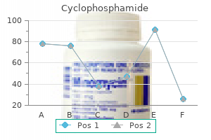
Buy cyclophosphamide 50 mg lowest price
Addition of escalating concentrations of ristocetin to platelet-rich plasma allows detection of each elevated and decreased sensitivity to ristocetin. Other methods to study platelet function embody circulate cytometry, electron microscopy, and next-generation sequencing. Flow cytometry has been used to research platelet construction and performance based on the detection of cell floor proteins with fluorescently labeled antibodies as nicely as detection of platelet activation. Electron microscopy can be used for the ultrastructural evaluation of platelets, together with evaluation of platelet granules and mobile organelles. Genomic approaches can additionally be used to determine specific inherited platelet defects, and next-generation sequencing is turning into more out there for prognosis of platelet disorders (Table 2. Third-step exams would include testing obtainable in specialized facilities, corresponding to biochemical studies, receptor binding assays, and molecular genetics research. However, as a lot as one third of both hemophilia A and hemophilia B circumstances may be the result of a spontaneous mutation, and there shall be no family historical past of the disease. Hemophilia A is the most typical inherited bleeding disorder, occurring in 1 in 5000 male births. In both ailments, the type and frequency of bleeding are determined by the degrees of the deficient issue. The most severe bleeding threat with severe illness happens early, with 1% to 4% of neonates experiencing intracranial hemorrhage, probably causing everlasting neurologic defects. Extracranial hemorrhage can even happen in severely affected neonates; each of these penalties are associated with traumatic supply. Severe hemophiliacs also undergo from spontaneous (not related to trauma) hemorrhage into muscle tissue and joints. The former usually happen in the muscular tissues of the legs, buttocks, and forearms and through the period when infants increase their exercise at 6 to eight months of age. Hemarthroses proceed to be an issue all through the life of a severe hemophiliac, increasing with age, and are a serious reason for morbidity in these patients. Repeated episodes of bleeding into a joint cause irritation, which finally damages the tissues within the joint causing hemophilic arthropathy and eventually joint deformity, permanent impairment, and ache. Moderate hemophilia is identified in older sufferers and is clinically heterogeneous. Although most patients expertise bleeding only after surgery or trauma, some may have spontaneous bleeding just like that seen in severely affected sufferers. These patients tend to be identified later in life, sometimes on routine coagulation screening. Female carriers of hemophilia are inclined to have factor levels round 50% and should experience menorrhagia, traumatic hemarthroses, bruising, hematomas, and postsurgical/postpartum bleeding. Risk of hemarthroses will increase with age, and hemophilic arthropathy happens in approximately half of sufferers with severe disease. In addition, some hemophiliacs who even have inherited thrombophilia, corresponding to issue V Leiden, might have fewer bleeds. The mainstay of remedy is intravenous issue alternative, normally with recombinant proteins, though in some nations purified plasma-derived products are still used. This could be given "in demand" as remedy for bleeding episodes or prophylactically in patients with a high danger for spontaneous bleeding. Prophylactic issue replacement has been shown to reduce joint harm in patients who exhibit hemarthroses. The earlier the patient begins prophylaxis, each in terms of age and length of time after first bleed, the higher the profit, and prophylaxis is commonly began at 2 years of age. Development of particular factor inhibitors, a threat for all patients with hemophilia, is mentioned further within the subsequent section. Antifibrinolytics, such as tranexamic acid and aminocaproic acid, can also assist with oral bleeding and epistaxis. Gene remedy can be an attractive technique, particularly since even a small increase in factor levels can have a significant impact on bleeding danger. Genetic analysis is really helpful in sufferers as a result of different types of gene defects are related to totally different levels of danger for developing inhibitors.
Discount 50 mg cyclophosphamide
The trigger is uncertain however is most probably related to irregular follicular dendritic cells. Each follicle may include one or more germinal middle; some follicles comprise no recognizable germinal heart. The germinal facilities include an increased proportion of follicular dendritic cells and endothelial cells. Mantle zone lymphocytes are organized in concentric rings ("onion skin" pattern) around the germinal center. Large, hyperchromatic, often bizarre, dystrophic cells with scant cytoplasm, in maintaining with irregular follicular dendritic cells, are typically found in the follicles, normally within the atrophic follicle centers, and less usually in the mantles. Lymphocytes are fewer in number than in a traditional lymph node, and there could also be an admixture of scattered plasma cells, few immunoblasts, few eosinophils, and clusters of plasmacytoid dendritic cells. Hyaline-vascular Castleman illness is usually an incidental discovering on radiographic examination, however patients may present with symptoms associated to compression of adjacent buildings or rarely with pain or a palpable mass. Patients have lymphadenopathy in a single website in almost all cases; in rare circumstances, the lesion entails an extranodal web site. A, Low energy reveals a lymph node replaced by follicles with broad mantles, inconspicuous follicle facilities, and a hypervascular interfollicular area. B, Higher power shows a follicle consisting mainly of small lymphocytes, with a suggestion of tiny sclerotic follicle centers, surrounded by numerous blood vessels. A, the enlarged lymph node shows areas with changes in maintaining with hyaline-vascular Castleman illness; two hyaline-vascular follicles are seen here. C, High energy shows spindle cells with oval to slightly elongate nuclei, vesicular chromatin, small nucleoli, and vague cytoplasmic borders. Follicular dendritic cells have aberrant expression of adhesion molecules and should present cytogenetic abnormalities. They may be asymptomatic, however systemic signs, together with fever, weight reduction, and fatigue, are frequent, and lots of have anemia, elevated erythrocyte sedimentation fee, and hypergammaglobulinemia. Following excision of the mass, the signs and laboratory abnormalities disappear, and sufferers do nicely. A, the lymph node exhibits a reactive follicle and sheets of interfollicular plasma cells. B, Higher power of the interfollicular area reveals a monotonous inhabitants of plasma cells. C and D, In this case, plasma cells categorical monotypic immunoglobulin (Ig), IgG sort (C,; D,). The mass lesion is composed of one enlarged lymph node or an combination of enlarged nodes, measuring about 3 to 11 cm, with individual nodes measuring 2. There are sheets of mature plasma cells in the interfollicular space, typically with Russell bodies; occasional immunoblasts may be seen. The follicles may be hyperplastic follicles of the same old sort or hyaline-vascular follicles or a combination of the two forms of follicles. In some cases (which might characterize an early stage of disease), immunoblasts and excessive endothelial venules, along with plasma cells, are current in massive numbers within the interfollicular region. The peak onset is within the fifth to sixth decade of life; these patients are youthful overall than in other cases of myeloma. The mild chain expressed is usually restricted to the V1 subfamily with a sample of somatic hypermutation suggesting antigen-driven choice. Other manifestations embody papilledema and evidence of extravascular quantity overload. A extensive variety of specific diseases could additionally be associated with morphologic options resembling these of Castleman disease, and these must be ruled out before making a diagnosis of Castleman illness. A, Low energy reveals a lymph node with a quantity of hyaline-vascular follicles and an interfollicular area with increased numbers of blood vessels. B, High energy of a portion of a follicle reveals a mantle zone composed of small lymphocytes with scattered massive lymphoid cells. A small space of follicle heart is seen throughout the mantle, and some blood vessels are seen in the interfollicular space on the alternative side of the mantle. Rare sufferers with systemic lupus erythematosus show lymphadenopathy with features of Castleman disease. Polytypic plasma cells exclude plasmacytoma however are often seen in plasma cell Castleman disease.
50 mg cyclophosphamide overnight delivery
Cytoplasm is scant, however visible, particularly on Giemsa staining, by which it has a basophilic high quality. In some instances, polylobated nuclei predominate as opposed to the everyday round contours. The occurrence of polylobated nuclei usually imparts a smaller cell measurement and might make distinction from small B-cell lymphomas tough. Multilobated cells are frequent in some extranodal lymphomas, similar to major lymphoma of bone. The lymphoma cells have vesicular chromatin and reasonable quantities of eosinophilic cytoplasm. Diagnosis of the immunoblastic variant is suitable when more than 90% of cells are of this kind. The anaplastic variant is composed of sheets of anaplastic, atypical cells with weird multilobated�multinucleated cells which will resemble Reed-Sternberg cells. Pleomorphic may be a greater term than anaplastic, as a result of the latter may be confused with anaplastic large-cell lymphoma of T-cell lineage. The diffuse sample is unusual and is normally seen when the lymphomatous infiltrate consists of large cells. The cytology is usually discordant (approximately half of the cases), that means the marrow infiltrate consists of small lymphocytes or blended small and large lymphocytes. Studies have shown that patients with concordant giant cells within the bone marrow have a poor response to remedy and poor overall survival. The cell-of-origin distinctions also have underpinnings in biology, with molecular genetics and next-generation sequencing having been useful in furthering our understanding of the molecular alterations underlying these disease subtypes. It is a loss-of-function mutation that forestalls terminal differentiation of the malignant B cells. Therefore consideration of morphologic, immunophenotypic, and molecular genetic options is required for correct analysis. Expression of B-cell markers and lack of markers anticipated in these different nonlymphoid malignancies, corresponding to keratins or melanomaassociated antigens, will help in appropriate analysis. Identification of these extra prognostic factors could assist decide alternative therapies in the future. Extranodal illness is common, with the spleen, liver, and bone marrow being the most commonly concerned extranodal websites. Malignant cells are scattered singly among the mobile background, though in some instances they might cluster. Some instances might have cells which have more standard immunoblastic or centroblastic features. Splenic involvement occurs in a multifocal and micronodular sample with enlargement of the white pulp by an infiltrate of cells comparable in composition to that described within the lymph node. Within the liver the lymphomatous infiltrate is confined principally to the portal tracts. Low magnification reveals predominance of small lymphocytes, scattered histiocytes, and occasional atypical large cells that represent the malignant cells (arrows). Patients could have headache, mental status modifications, or focal neurologic signs associated to mass impact at the web site of involvement. In one collection, 81% of circumstances occurred in a supratentorial location, 7% infratentorially, and 12% in both areas. Because this neoplasm grows rapidly, the length of signs and signs is on the order of weeks. Given the perivascular association of lymphoid cells, inflammatory situations similar to encephalitis should be considered. Comparative genomic hybridization research have proven that gains outnumber losses, with chromosome 12q showing the most frequent positive aspects (63%). The mostly deleted locus is situated at chromosome 6q (47%), with 6q21-22 being the commonly deleted area. The latter is 282 typically withheld in older sufferers because of unacceptable toxicity.
Real Experiences: Customer Reviews on Cyclophosphamide
Irmak, 27 years: This smear from a affected person with severe -thalassemia demonstrates erythroblastosis, outstanding hypochromia, target cells, and other weird poikilocytes. B, the pale areas correspond to quite a few antigen-presenting cells, including Langerhans cells with pale, folded, and grooved nuclei.
Sibur-Narad, 24 years: Frog leg lateral radiograph of both hips demonstrating a left-sided Investigations 7 1. Clitoral e nlargemem witJ1 ambiguo us genitalia at b irth may be d ue to feminine pseudohermaphroditism, blended gonadal dysgenesis, male pseudohermaphroditism and t-arel) true he nnaphroditism.
Ilja, 47 years: Surgical expertise in hip arthroscopy are additionally growing, and that is certain to improve cartilage restoration techniques regardless of the cells getting used. The extra Osteotomies [1, 8] proximal the level of osteotomy, the greater the attainable correction, so these are usually reserved for higher deformities.
Vak, 23 years: Treatment with corticosteroids and alcoholism or substance abuse are additionally known to be associated with elevated threat of fracture [28]. Dyspoietic modifications may be recognized in different circumstances with florid erythroid hyperplasia.
Kamak, 42 years: Jaiswal S, Fontanillas P, Flannick J, et al: Age-related clonal hematopoiesis related to adverse outcomes, N Engl J Med 371:2488�2498, 2014. Witl1 tl1e passage of time, the frequency and sevetity of flushes climinish over a interval of 1-2 years.
9 of 10 - Review by V. Jose
Votes: 164 votes
Total customer reviews: 164
References
- Cramer SC, Hill MD. Human choriogonadotropin and epoetin alfa in acute ischemic stroke patients (REGENESIS-LED trial). Int J Stroke 2014;9(3):321-7.
- Bosworth BM. Calcium deposits in the shoulder and subacromial bursitis: a survey of 12,122 shoulders. JAMA 1941; 116:2477-82.
- Straumsheim P, Borchgrevink C, Mowinkel P, et al. Homoepathic treatment of migraine: a double blind, placebo controlled trial of 68 patients. Br Homeopath J. 2000;89:4-7.
- Gladding PA, Webster MW, Kay P. Late drug-eluting stent thrombosis and erythropoietin: cause and effect? Heart Lung Circ 2007;16:305-307.
- Skarin AT, Herbst RS, Leong TL, Bailey A, Sugarbaker D. Lung cancer in patients under age 40.
- Gupta M, Haluck RS, Yang HC, et al. Laparoscopic-assisted renal biopsy: an alternative to open approach. Am J Kidney Dis. 2000;36(3):636-639.
- Pogrel MA. Evaluation of over 400 autogenous tooth transplants. J Oral Maxillofac Surg 1987;45:205-211.
- Schellinger PD, Meinck HM, Thron A. Diagnostic accuracy of MRI compared to CCT in patients with brain metastases. J Neurooncol 1999;44(3):275-281.