Malegra FXT Plus
Malegra FXT Plus dosages: 160 mg
Malegra FXT Plus packs: 20 pills, 30 pills, 60 pills, 90 pills, 120 pills, 180 pills, 270 pills
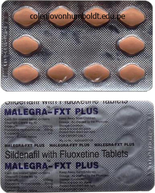
Buy malegra fxt plus with american express
The plateau is a sustained interval of depolarization, which accounts for the lengthy length of the action potential and, consequently, the long refractory durations. An motion potential in a Purkinje fiber (not shown) would look much like that in the ventricular fiber, but its length could be barely longer. In ventricular, atrial, and Purkinje fibers, the motion potential begins with a phase of rapid depolarization, called the upstroke. As in nerve and skeletal muscle, the upstroke is brought on by a transient enhance in Na+ conductance (gNa), produced by depolarization-induced opening of activation gates on the Na+ channels. At the height of the upstroke, the membrane potential is depolarized to a value of about +20 mV. Thus dV/dThis best (the fee of rise of the upstroke is fastest) when the resting membrane potential is most negative, or hyperpolarized. This correlation relies on the connection between membrane potential and the position of the inactivation gates on the Na+ channel (see Chapter 1). Phase 1 in ventricular, atrial, and Purkinje fibers is a quick interval of repolarization, which immediately follows the upstroke. There are two explanations for the occurrence of the web outward current during phase 1. First, the inactivation gates on the Na+ channels close in response to depolarization. When these gates shut, gNa decreases and the inward Na+ present (which triggered the upstroke) ceases. Because the K+ 136 � Physiology conductance (gK) is high, K+ flows out of the cell, down this steep electrochemical gradient. There is an increase in calcium (Ca2+) conductance (gCa), which leads to an inward Ca2+ present. Inward Ca2+ present can additionally be called slow inward current, reflecting the slower kinetics of those channels (compared with the fast Na+ channels of the upstroke). The Ca2+ channels that open in the course of the plateau are L-type channels and are inhibited by the Ca2+ channel blockers nifedipine, diltiazem, and verapamil. Thus during the plateau, the inward Ca2+ current is balanced by the outward K+ present, the online current is zero, and the membrane potential remains at a stable depolarized value. This Ca2+ entry in the course of the plateau of the motion potential initiates the release of extra Ca2+ from intracellular stores for excitation-contraction coupling. This strategy of so-called Ca2+-induced Ca2+ launch is discussed within the part on cardiac muscle contraction. Recall that repolarization is produced when outward currents are greater than inward currents. During phase three, repolarization outcomes from a combination of a lower in gCa (previously increased in the course of the plateau) and an increase in gK (to even larger levels than at rest). At the end of phase three, the outward K+ present is reduced as a end result of repolarization brings the membrane potential closer to the K+ equilibrium potential, thus decreasing the driving force on K+. The membrane potential absolutely repolarizes throughout phase 3 and returns to the resting level of roughly -85 mV. During section four, the membrane potential is stable once more, and inward and outward currents are equal. The K+ channels, and the resulting K+ present, liable for phase 4 are different from these responsible for repolarization in section three. The steady membrane potential in section 4 signifies that inward and outward currents are equal. The query might arise: How can the sum of inward Na+ and Ca2+ currents be the same magnitude because the outward K+ current, given that gNa and gCa are very low and gK1 is very excessive The answer lies in the truth that, for each ion, present = conductance � driving drive. Although gK1 is excessive, the driving drive on K+ is low because the resting membrane potential is near the K+ equilibrium potential; thus the outward K+ current is relatively small. On the other hand, gNa and gCa are each low, however the driving forces on Na+ and Ca2+ are excessive as a end result of the resting membrane potential is way from the Na+ and Ca2+ equilibrium potentials; thus the sum of the inward currents carried by Na+ and Ca2+ is equal to the outward present carried by K+. In the opposite myocardial cells, the upstroke is the results of an increase in gNa and an inward Na+ present. The "f," which stands for funny, denotes that this Na+ current differs from the fast Na+ present answerable for the upstroke in ventricular cells. Once If and sluggish depolarization bring the membrane potential to threshold, the Ca2+ channels are opened for the upstroke.
Malegra fxt plus 160mg online
As the neural plate curves over to form the neural tube, the previous apical surface is situated surrounding the lumen (the future ventricles), while the previous basal floor turns into the outer surface of the neural tube positioned just below the pial membrane. Sauer reported that cells positioned close to the ventricular surface lengthen skinny processes that contact both the apical and basal surfaces. The nuclei of these cells then travel via these processes as the cell cycle progresses. As the neural plate curves over to kind the neural tube, the apical surface of the neural plate turns into the ventricular surface surrounding the lumen of the neural tube and the basal surface becomes the outer (pial) surface. Sauer was the first to report that the cells along the ventricular floor prolong cytoplasmic processes that contact both the pial and ventricular floor. The nucleus of the cell then migrates through the cytoplasmic process utilizing a mechanism referred to as interkinetic nuclear migration. The nucleus first travels to the basal (pial) floor, then returns to the apical (ventricular) floor, the place it undergoes mitosis (cell division). The motion of the nucleus via the cytoplasmic process was referred to as intermitotic migration of nuclei or, as is usually referred to as at present, interkinetic nuclear migration. In the Nineteen Fifties she and colleagues conducted a number of experiments using nuclear stains and the newly developed tritiated thymidine technique that confirmed nuclear migration was linked to phases of the cell cycle. Once chromosomes are copied, the cells progress to the G2 (gap 2) stage, preparing for mitosis. Sauer reported the sample of nuclear migration in neural precursor cells within the neural tube of the chick. This report was one of the first neurobiology experiments to use the tritiated thymidine labeling method developed in 1957. Thus, nuclei of cells on the G1 phase of the cell cycle are positioned on the ventricular zone. When the migrating nucleus reaches the pial surface, the cell is in the S section of the cell cycle, after which the nucleus travels back to the ventricular floor, the place the cell enters the M part and divides into two dn 5. The resulting daughter cells will either migrate away from the ventricular floor, or prolong a course of to the pial floor and continue by way of another series of interkinetic actions so as to divide once more. As development progresses and more precursor cells are generated, the wall of the neural tube thickens so that the cell nuclei should journey more and more higher distances to the pial surface. It is thought that the nucleus may be exposed to development factors or other signals within the cytoplasm because it travels by way of the completely different areas of the cell course of in the course of the numerous stages of the cell cycle. It has additionally been advised that the movement of nuclei into different layers provides extra space for proliferation to happen in the very small area of the early neural tube. These unique and really exactly ordered patterns of interkinetic motion are seen only in areas of pseudostratified epithelium and the ventricular zone of the neural tube. Thus, the intriguing strategy of interkinetic actions is restricted to particular areas of the creating embryo. The last stage is the M part (mitosis) by which the cell divides to produce two daughter cells. Sauer demonstrated that cells that incorporated a radiolabel in the course of the S part of the cell cycle were positioned near the pial (basal) surface of the neural tube. Sauer confirmed that the translocation of the nucleus corresponds to totally different levels of the cell cycle. The cell enters the gap 1 (G1) section of the cell cycle as its nucleus travels towards the pial (basal) floor. Once the nucleus reaches the ventricular (apical) surface, it enters the mitotic (M) phase during which the cell divides. The plane of cell division and patterns of protein distribution decide whether or not a cell proliferates or migrates Following interkinetic translocation of the nucleus and its return to the ventricular surface, the precursor cell divides. The cleavage airplane of cell division is crucial in figuring out whether or not a cell continues to divide or migrates away from the ventricular zone. Cell division happens alongside a vertical cleavage aircraft (perpendicular to the ventricular surface) or either a horizontal (parallel to the ventricular surface) or an oblique cleavage plane (at an angle to the ventricular surface).
Diseases
- Katz syndrome
- Cantalamessa Baldini Ambrosi syndrome
- Glanzmann thrombasthenia
- Meretoja syndrome
- Esophageal atresia coloboma talipes
- Rosenberg Lohr syndrome
- Marie Unna congenital hypotrichosis
- Urticaria-deafness-amyloidosis
- Chromosome 19, trisomy 19q
Order 160 mg malegra fxt plus visa
The serous cells secrete an aqueous fluid, and the mucous cells secrete mucin glycoproteins for lubrication. Initial saliva is produced by acinar cells (1) and subsequently modified by ductal epithelial cells (2). The acinar cells produce an preliminary saliva composed of water, ions, enzymes, and mucus. This preliminary saliva passes through a brief phase, referred to as an intercalated duct, after which via a striated duct, which is lined with ductal cells. The ductal cells modify the preliminary saliva to produce the final saliva by altering the concentrations of varied electrolytes. When stimulated by neural input, the myoepithelial cells contract to eject saliva into the mouth. Salivary acinar cells and ductal cells have both parasympathetic and sympathetic innervation. Although many organs have such dual innervation, the weird feature of the salivary glands is that saliva production is stimulated by each the parasympathetic and the sympathetic nervous systems (although parasympathetic management is dominant). The salivary glands have an unusually excessive blood move that increases when saliva production is stimulated. When corrected for organ dimension, maximal blood move to the salivary glands is more than 10 instances the blood move to exercising skeletal muscle! Formation of Saliva Saliva is an aqueous solution whose volume may be very high contemplating the small size of the glands. Saliva consists of water, electrolytes, -amylase, lingual lipase, kallikrein, and mucus. The first step is the formation of an isotonic plasma-like resolution by the acinar cells. The acinar cells secrete the initial saliva, which is isotonic and has approximately the same electrolyte composition as plasma. The transport mechanisms involved on this modification are complicated, but they are often simplified by considering events within the luminal and basolateral membranes individually and then by figuring out the web result of all of the transport mechanisms. A last query is How does saliva, which was initially isotonic, turn into hypotonic as it flows via the ducts The acinar cells additionally secrete organic constituents corresponding to -amylase, lingual lipase, mucin glycoproteins, IgA (immunoglobulin A), and kallikrein. Kallikrein is an enzyme that cleaves high-molecular-weight kininogen into bradykinin, a potent vasodilator. During intervals of high-salivary gland activity, kallikrein is secreted and produces bradykinin. Bradykinin then causes local vasodilation, which accounts for the highsalivary blood flow during times of elevated salivary exercise. At the highest move charges (4 mL/min), the final saliva most intently resembles plasma and the preliminary saliva produced by the acinar cells. At the lowest move charges (<1 mL/min), the ultimate saliva is most dissimilar to plasma (it has lower concentrations of Na+ and Cl- and a better focus of K+). The mechanism of the flow-rate�dependent modifications in focus relies totally on the period of time saliva is in contact with the ductal cells. Parasympathetic activity to the salivary glands is increased by food, scent, and nausea and by conditioned reflexes. The sympathetic enter to the salivary glands originates in thoracic segments T1�T3 with preganglionic nerves that synapse in the superior cervical ganglion. The postganglionic sympathetic neurons launch norepinephrine, which interacts with -adrenergic receptors on the acinar and ductal cells. Sympathetic stimulation also prompts -adrenergic receptors on acinar cells, although the activation of -adrenergic receptors is taken into account more essential. Gastric Secretion the cells of the gastric mucosa secrete a fluid known as gastric juice. Structure and Cell Types of the Gastric Mucosa the anatomic divisions of the stomach (fundus, body, and antrum) have been mentioned in the section on motility.
Purchase 160mg malegra fxt plus
Because thyroid hormones enhance O2 consumption, they create the next demand for O2 within the tissues. Increased O2 supply to the tissues is feasible as a outcome of thyroid hormones produce a rise in cardiac output and air flow. The improve in cardiac output is the result of a combination of increased heart price and elevated stroke volume (increased contractility). These cardiac effects are defined by the fact that thyroid hormones induce the synthesis of. Recall that these 1 receptors mediate the results of the sympathetic nervous system to increase heart fee and contractility. Thus when thyroid hormone ranges are high, the myocardium has an elevated number of 1 receptors and is extra delicate to stimulation by the sympathetic nervous system. Thyroid hormones act synergistically with development hormone and somatomedins to promote bone formation. Thyroid hormones promote ossification and fusion of bone plates and bone maturation. In adults, hypothyroidism causes listlessness, slowed movement, somnolence, impaired reminiscence, and decreased mental capacity. The results of thyroid hormones and catecholamines on warmth production, cardiac output, lipolysis, and gluconeogenesis seem to be synergistic. The significance of this synergism is illustrated by the effectiveness of -adrenergic blocking brokers. Pathophysiology of Thyroid Hormone the most common endocrine abnormalities are disturbances of thyroid hormones. Hyperthyroidism the most common form of hyperthyroidism is Graves illness, an autoimmune disorder characterized by increased circulating levels of thyroid-stimulating immunoglobulins. When current, the antibodies intensely stimulate the thyroid gland, resulting in increased secretion of thyroid hormones and hypertrophy of the gland. The analysis of hyperthyroidism is predicated on signs and measurement of elevated ranges of T3 and T4. Treatment of hyperthyroidism consists of administration of medication similar to propylthiouracil, which inhibit the synthesis of thyroid hormones; surgical elimination of the gland; or radioactive ablation of the thyroid gland with 131I-. Hypothyroidism the most common explanation for hypothyroidism is autoimmune destruction of the thyroid gland (thyroiditis) 9-Endocrine Physiology � 427 in which antibodies may both frankly destroy the gland or block thyroid hormone synthesis. Other causes of hypothyroidism are surgical elimination of the thyroid as remedy for hyperthyroidism, hypothalamic or pituitary failure, and I- deficiency. Rarely, hypothyroidism is the result of target tissue resistance attributable to down-regulation of thyroid-hormone receptors. The prognosis of hypothyroidism is based on signs and a discovering of decreased ranges of T3 and T4. The symptoms of hypothyroidism are opposite those seen in hyperthyroidism and embody decreased metabolic rate and weight achieve without elevated meals consumption; decreased heat manufacturing and chilly intolerance; decreased heart price; slowing of movement, slurred speech, slowed psychological exercise, lethargy, and somnolence; periorbital puffiness; constipation; hair loss; and menstrual dysfunction. Finally, and of critical importance, if hypothyroidism happens in the perinatal interval and is untreated, it leads to an irreversible form of progress and mental retardation known as cretinism. Treatment of hypothyroidism entails thyroid hormone substitute therapy, often T4. Like endogenous hormone, exogenous T4 is converted to its energetic form, T3, within the target tissues. In Graves disease, the most common reason for hyperthyroidism, the high levels of thyroidstimulating immunoglobulins drive extra secretion of T4 and T3 and nonetheless have a trophic effect on the thyroid gland to produce goiter. Autoimmune thyroiditis is a standard reason for hypothyroidism, by which thyroid hormone synthesis is impaired by antibodies to peroxidase, leading to decreased T4 and T3 secretion. The terms hyperthyroid, hypothyroid, and euthyroid describe, respectively, the medical states of extra thyroid hormone, deficiency of thyroid hormone, and normal ranges of thyroid hormone. Thus they describe blood levels of thyroid hormone, not the scale of the thyroid gland. The presence or absence of goiter could be understood solely by analyzing the etiology of the varied thyroid disorders. The adrenal glands are literally two separate glands, the adrenal medulla and the adrenal cortex, whose secretions are essential for all times. The adrenal medulla, which is within the inner zone of the gland, composes roughly 20% of the tissue.
Buy 160 mg malegra fxt plus
However, the micturition reflex itself is managed by the autonomic nervous system. The sympathetic innervation of the detrusor muscle and the interior sphincter originates within the lumbar spinal twine (L1�L3), and the parasympathetic innervation originates within the sacral spinal twine (S2�S4). This sympathetic activity produces relaxation of the detrusor muscle, through 2 receptors, and contraction of the inner sphincter muscle, by way of 1 receptors. When the muscle wall is relaxed and the sphincters are closed, the bladder can fill with urine. When the bladder is full, this fullness is sensed by mechanoreceptors within the bladder wall, and afferent neurons transmit this info to the spinal cord and then to the mind stem. The micturition reflex is coordinated by centers within the midbrain, and now para sympathetic management predominates. Parasympathetic exercise produces contraction of the detrusor muscle (to improve pressure and eject urine) and rest of the inner sphincters. Clearly, the sympathetic and parasympathetic actions on the bladder constructions are reverse but coordinated: the sympathetic actions dominate for bladder filling, and the parasympathetic actions dominate for bladder emptying. The pupillary dilator muscle is managed by sympathetic innervation via 1 receptors. Activation of those 1 receptors causes constriction of the radial muscle, which causes dilation of the pupil, or mydriasis. The pupillary constrictor muscle is controlled by parasympathetic innervation via muscarinic receptors. During filling of the bladder, sympathetic control predominates, causing leisure of the detrusor muscle and contraction of the internal sphincter. During micturition, parasympathetic control predominates, inflicting contraction of the detrusor muscle and rest of the inner sphincter. Dashed lines symbolize sympathetic innervation; stable traces symbolize parasympathetic innervation. Activation of those muscarinic receptors causes con striction of the sphincter muscle, which causes con striction of the pupil, or miosis. In the lodging response, a blurred retinal picture activates parasympathetic preganglionic neurons within the Edinger-Westphal nuclei and leads to contraction of the sphincter muscle and pupillary constriction. At the same time, the ciliary muscle contracts, causing the lens to "spherical up" and its refractive power to improve. There are some notable exceptions to the generalization of reciprocal innervation. Several organs have only sympathetic innervation: sweat glands, vascular clean muscle, pilomotor muscular tissues of the pores and skin, liver, adipose tissue, and kidney. Thus sympathetic exercise dominates when the bladder is filling to produce relaxation of the bladder wall and, simultaneously, contraction of the interior bladder sphincter. The bladder can fill as a outcome of the bladder wall is relaxed and the sphincter is closed. During micturition, parasympathetic exercise dominates, producing contraction of the bladder wall and, simultaneously, leisure of the sphincter. Similar reasoning can be applied to the autonomic management of the gastrointestinal tract: Contraction of the wall of the gastrointestinal tract is accompanied by rest of the sphincters (parasympathetic), allowing the contents of the gastrointestinal tract to be propelled forward. Relaxation of the wall of the gastrointestinal tract is accompanied by contraction of the sphincters (sympathetic); the combined effect of these actions is to gradual or stop motion of the contents. Types of Receptors Coordination of operate throughout the organ techniques, as orchestrated by the autonomic nervous system, is one other recurring physiologic theme (Boxes 2. This management is exquisitely clear, for instance, when contemplating the operate of the urinary bladder. These generalizations are as follows: (1) In the parasympathetic division, effector organs have muscarinic receptors. A 66-year-old man who suffered a stroke on the right facet has a drooping proper eyelid (ptosis), constriction of his right pupil (miosis), and lack of sweating on the best facet of his face (anhidrosis). When a solution of 10% cocaine is applied in the left eye, it causes dilation of the pupil (mydriasis).
Syndromes
- Inability to fully extend the joints from birth (contracture deformity)
- Do not use creams or other medicines in the vagina.
- Has bluish skin
- Cough
- CIN II -- moderate to marked dysplasia
- Hearing loss
- Creatinine clearance - blood and urine
- Fluids through a vein (IV)
- The machine may move your head so that the energy beams are delivered to the exact spots that need treatment.
- Bone scan
Cheap malegra fxt plus 160 mg
Energy: Low vitality and/or fatigue are frequent complaints in melancholy, as is problem in getting started or initiating tasks. The fatigue skilled could be physical or mental, and could also be related to poor sleep and appetite. In extreme instances, routine actions such as every day hygiene, grooming, or consuming could additionally be impaired. Depressed patients may misread trivial every day events and take accountability for adverse occasions out of their control; these can generally be of delusional proportion. Concentration: Difficulty with focus and decision-making is often experienced in melancholy. In elderly sufferers, the cognitive complaints may be misdiagnosed as early dementia. Overeating, accompanied by decreased exercise and exercise, can lead to weight gain and metabolic syndrome. Psychomotor retardation consists of slowing (slowed physique movements, lack of facial features, long latency of speech response) which, at its extreme, can manifest as mute or catatonic shows. Anxiety can also present as psychomotor agitation (talking shortly, pacing, restlessness, incapability to sit still). Suicide: Some sort of suicidal ideation, ranging from fleeting ideas of wishing everything would finish to elaborate plans for suicide, is present in almost two-thirds of people with depression. Even when suicidal ideas are serious, depressed patients typically lack the energy and motivation to attempt suicide. However, suicide stays a major issue as 0�5% of hospitalized depressed people ultimately die by suicide. A period of high danger for suicide is during preliminary remedy, when power and motivation might enhance earlier than the cognitive symptoms. Other symptoms: Although not formally indicated as criteria for the analysis, a variety of other signs and indicators, including nervousness, irritability, cognitive dysfunction, and pain, are related to despair. The diagnostic criteria require a threshold variety of symptoms that have to be current much of the time, most days, for no less than 2 weeks, although the period is often for a lot longer by the point that help is sought. The symptoms also must considerably impair functioning or cause important distress. No Yes Dysthymia (Persistent depressive disorder) Other speci ed depressive disorders Major depressive disorder with residual symptoms Yes Prior manic/ hypomanic episode Dysthymic signs can develop slowly, often unrecognized by the individual, and persist for a minimum of two years (median 5 years). Depressive problems that happen secondary to substance use and different medical situations are described in sections 4. In children, this may current as failure to obtain a typical weight for their age. Full symptom standards for a serious depressive episode could also be current during the 2 years. These behaviours are out of proportion to a situation, not according to the developmental stage, and not part of a hypomanic or manic episode. Premenstrual dysphoric dysfunction: In most menstrual cycles during the earlier year, signs frequently occurred over the last week of the luteal phase and remitted inside a couple of days of the onset of menses. Other specified depressive problems: these embody displays corresponding to minor melancholy (sub-syndromal episodes with inadequate symptoms), recurrent temporary depression, and short-episode. Minimal number of criterion symptoms with gentle distress and manageable intensity 2. Most criterion symptoms are current with severe distress and unmanageable depth 2. While these standards could additionally be of heuristic value, validated depression ranking scales are more clinically helpful for assessing severity (see Chapter 6 and Appendix). For example, psychotherapy is as effective as pharmacotherapy for mild-to-moderate depression, however severe despair shows higher response to mixture treatment. Emerging proof additionally suggests that some antidepressants could additionally be more effective than others for severe despair (see Chapter 8). Bereavement Bereavement or grief over lack of relationships can share similar signs. Evidence from the historical past, physical examination, or laboratory findings is used to set up whether abuse, dependence, intoxication, or withdrawal states are physiologically inducing a depressive episode.
Order line malegra fxt plus
Additionally, because the retinotectal system has been studied for so a few years, it is rather nicely characterised and thus offers scientists with a wealth of data on which to draw. The following sections describe the findings that first led scientists to examine axon-target recognition within the retinotectal system and the next experiments used to identify specific cues to regulate the proper mapping of retinal ganglion cell axons across the optic tectum. Several scientists at that time favored the concept that once axons managed to reach the target tissue, a selected target cell acknowledged and shaped a synaptic connection solely with an axon that supplied an identical sample of neural exercise. The notion of a target cell responding to the neural exercise of an axon was known as the resonance hypothesis-that is, the goal cells would resonate solely with axons offering matching electrical activity. This speculation was developed over a variety of years largely by way of the work of Paul Weiss and colleagues. Among the most pivotal experiments that reshaped how scientists think about axonal steerage mechanisms have been those carried out by Roger Sperry from the Forties to the Nineteen Sixties. Although Sperry worked with Weiss, he saw limits to the resonance speculation and so started a series of experiments using the retinotectal system of amphibians to test how axons acknowledge and make correct connections with target cells. A number of studies within the early 1900s revealed that if the optic nerve was crushed or severed, the retinal axons would regenerate and reestablish connections within the optic tectum. New nerve fibers had sprouted from the reduce stump and had managed to grow back to the visual facilities of the mind. And but, this was the one potential rationalization, for without question the newts had regained normal imaginative and prescient. This group also discovered that if an eye from one salamander was transplanted to one other salamander, the retinal axons of the transplanted eye regenerated and restored imaginative and prescient. Because vision was restored even after multiple surgical procedures, Sperry and others acknowledged that the retinotectal system would offer a way of testing how axonal connections are established with specific goal cells. Other scientists hypothesized that the axons regrew in a scientific method to reestablish their original connections with specifc goal cells. This latter speculation was consistent with the idea that a chemical cue directed the axons to a particular goal cell. To test whether or not retinal axons relied on target-derived, chemical cues to project to a selected area of the tectum, Sperry modified the attention surgical procedure technique used by Matthey and Stone. In his 1943 report, Sperry completed this surgical procedure on 58 adult newts and famous they recovered visual perform over a period ranging from 28�95 days. When the lure was introduced in front of its head, it might flip around and start searching in the rear; when the bait was behind it, the animal would lunge forward. Even those animals that survived for two years continued to behave as if the visual world have been rotated a hundred and eighty degrees. Experiments in frogs, toads, and fish revealed comparable outcomes following eye rotations. As in the newts, the visual world was inverted and no quantity of apply compensated for the inverted visible field. Sperry proposed a mechanism by which retinal axons put out quite a few branches and tested completely different cells till "ultimately the rising tip encounters a cell floor for which it has a selected chemical affinity and to which it adheres. Studies by Roger Sperry from the Nineteen Forties through the 1960s indicated that retinotectal projections returned to the unique target cells after experimental manipulations. After the attention was rotated, the optic nerve was reduce and the animal was allowed to recuperate. The giant arrows indicate the course of movement of an object, while the small arrows point out the course of head movement. The experimental animals all the time responded as if the object were moving in the reverse direction. In the strictest sense, chemoaffinity would require matching "chemical tags," as Sperry called them, between every axon and each goal cell. Retinotectal maps are found in regular and experimental circumstances To better understand the group of retinal axons inside the tectum, scientists mapped axonal connections utilizing histological preparations and electrophysiological recordings. This is one example of a topographic map-the consistent, primarily invariant projection of axons from one area of the nervous system onto another. The behavioral results obtained after the eye rotation surgeries indicated that the temporal axons should nonetheless develop to the anterior tectum and the nasal axons to the posterior tectum, even after these experimental manipulations. Throughout the Fifties, 1960s, and 1970s, scientists used a selection of strategies to additional evaluate the mapping of retinotectal projections under completely different experimental situations.
Order malegra fxt plus 160mg fast delivery
It means that the sample and timing of the electrical activation of the guts are normal. Concepts Associated With Cardiac Action Potentials the cell, which known as an inward present. The permeant ion then will move into or out of the cell in an try to reestablish electrochemical equilibrium, and this current flow will alter the membrane potential. For example, consider the effect of lowering the extracellular K+ focus on the resting membrane potential of a myocardial cell. The K+ equilibrium potential, calculated by the Nernst equation, will become more adverse. K+ ions will then move out of the cell and down the now larger electrochemical gradient, driving the resting membrane potential toward the new, more negative K+ equilibrium potential. For instance, the resting permeability of ventricular cells to Na+ is quite low, and Na+ contributes minimally to the resting membrane potential. However, during the upstroke of the ventricular motion potential, Na+ conductance dramatically increases, Na+ flows into the cell down its electrochemical gradient, and the membrane potential is briefly pushed toward the Na+ equilibrium potential. At threshold potential, the depolarization turns into selfsustained and gives rise to the upstroke of the action potential. Action Potentials of Ventricles, Atria, and the Purkinje System the concepts utilized to cardiac motion potentials are the same concepts that are applied to action potentials in nerve, skeletal muscle, and clean muscle. The following part is a abstract of these rules, which are discussed in Chapter 1: 1. The membrane potential of cardiac cells is decided by the relative conductances (or permeabilities) to ions and the concentration gradients for the permeant ions. If the cell membrane has a excessive conductance or permeability to an ion, that ion will move down its electrochemical gradient and try and drive the membrane potential towards its equilibrium potential (calculated by the Nernst equation). If the cell membrane has low conductance or permeability to an ion or is impermeable to the ion, that ion will make little or no contribution to the membrane potential. By conference, membrane potential is expressed in millivolts (mV), and intracellular potential is expressed relative to extracellular potential; for instance, a membrane potential of -85 mV means 85 mV, cell inside negative. The resting membrane potential of cardiac cells is determined primarily by potassium ions (K+). The conductance to K+ at rest is high, and the resting membrane potential is close to the K+ equilibrium potential. Since the conductance to sodium (Na+) at rest is low, Na+ contributes little to the resting membrane potential. Changes in membrane potential are caused by the move of ions into or out of the cell. The motion potential in these tissues shares the next traits (Table 4. Action potential length varies from a hundred and fifty ms in atria, to 250 ms in ventricles, to 300 ms in Purkinje fibers. These durations may be in contrast with the temporary period of the motion potential in nerve and skeletal muscle (1�2 ms). Recall that the length of the motion potential additionally determines the period of the refractory intervals: the longer the action potential, the longer the cell is refractory to firing another motion potential. The cells of the atria, ventricles, and Purkinje system exhibit a stable, or fixed, resting membrane potential. The action potential in cells of the atria, ventricles, and Purkinje system is characterised by a plateau. The results of the autonomic nervous system on coronary heart price are based on such adjustments in the fee of section four depolarization and are discussed later in the chapter. The rule is that the pacemaker with the quickest rate of phase 4 depolarization controls the heart fee. On the other hand, the rapid conduction velocity of the Purkinje fibers ensures that the ventricles may be activated rapidly and in a smooth sequence for efficient ejection of blood.
Discount 160mg malegra fxt plus amex
The cellular mechanism for this glucose reabsorption is the Na+glucose cotransporter in the luminal membrane of the proximal tubule cells. Once these binding sites are absolutely occupied, saturation of transport happens (transport maximum). Insulin is required for normal uptake of glucose into liver, muscle, and other cells. When the blood glucose concentration increases to high ranges, extra glucose is filtered by the renal glomeruli and the amount of glucose filtered exceeds the capacity of the Na+-glucose cotransporter. Treatment of the patient with type I diabetes mellitus consists of administering exogenous insulin by injection. Whether secreted usually from the pancreatic cells or administered by injection, insulin lowers the blood glucose concentration by promoting glucose uptake into cells. When this patient acquired insulin, his blood glucose concentration was decreased; thus the quantity of glucose filtered was reduced, and the Na+-glucose cotransporters had been now not saturated. All of the filtered glucose could be reabsorbed, and due to this fact no glucose was excreted, or "spilled," in the urine. To accomplish Ca2+-Na+ change, energetic transport should be involved as a outcome of Ca2+ moves out of the cell against its electrochemical gradient. The protein should bind Ca2+ on the intracellular facet of the membrane and, concurrently, bind Na+ on the extracellular facet. In this configuration, the change protein rotates and delivers Ca2+ to the exterior of the cell and Na+ to the interior of the cell. The stoichiometry of Ca2+-Na+ trade varies between completely different cell types and may even range for a single cell kind under different circumstances. Usually, nonetheless, three Na+ ions enter the cell for every Ca2+ ion extruded from the cell. With this stoichiometry of three Na+ ions per one Ca2+ ion, three optimistic charges move into the cell in trade for two constructive costs leaving the cell, making the Ca2+-Na+ exchanger electrogenic. Osmosis Osmosis is the flow of water throughout a semipermeable membrane because of variations in solute focus. Concentration differences of impermeant solutes establish osmotic pressure differences, and this osmotic pressure distinction causes water to circulate by osmosis. If two solutions have totally different calculated osmolarities, the solution with the higher osmolarity known as hyperosmotic and the solution with the decrease osmolarity is identified as hyposmotic. Solution A has the next osmolarity than Solution B and is hyperosmotic; Solution B is hyposmotic. Because 1 kg of water is roughly equal to 1 L of water, osmolarity and osmolality will have primarily the same numerical value. Osmosis is the circulate of water throughout a semipermeable membrane as a end result of a difference in solute focus. The difference in solute concentration creates an osmotic strain distinction throughout the membrane and that strain distinction is the driving drive for osmotic water move. The membrane separating the options is permeable to water however is impermeable to the solute. The solute in Solution 1 produces an osmotic pressure and causes, by the interplay of solute with pores in the membrane, a discount in hydrostatic strain of Solution 1. The ensuing hydrostatic strain difference throughout the membrane then causes water to move from Solution 2 into Solution 1. With time, water move causes the amount of Solution 1 to increase and the volume of Solution 2 to decrease. The stress required to stop the circulate of water is the osmotic pressure of Solution 1. The osmotic strain of Solution 1 is dependent upon two factors: the concentration of osmotically active particles and whether the solute stays in Solution 1. Solution B accommodates NaCl, which dissociates partially in resolution however not fully. A, Solute (circles) is present on one facet of a semipermeable membrane; with time, the osmotic pressure created by the solute causes water to circulate from Solution 2 to Solution 1.
Purchase malegra fxt plus overnight delivery
This discovering suggested there have been additionally intrinsic temporal cues present to affect fate options of cortical neurons. A third set of experiments confirmed that the destiny potential of the cortical neurons becomes gradually restricted throughout regular improvement. Together, the transplantation experiments revealed early-mid-stage progenitors are multipotent early within the cell cycle and might adopt new cortical fates when positioned in an older host surroundings. As cells reach mid-late phases of development they turn out to be restricted in cell fate choices. Thus, the progenitors arising from mid-late levels of cortical development are unable to undertake the fates of youthful progenitors and remain committed to the fate similar to their time of migration. A variety of subsequent studies have identified transcription components which are specific to neurons situated in several cortical layers. Thus, early-stage cortical progenitors are able to alter their fate and migrate to a new layer. However, this impact was solely seen if the cells were harvested early within the cell cycle, previous to the final mitotic division. It has been advised that some mixtures of transcription components are mutually repressive to prevent cells from adopting the destiny of cells in adjacent layers. Ongoing research seek to identify how transcription elements determine cortical layer cell destiny. In a minimal of some circumstances, the recognized transcription factors regulate differentiation of neurons as soon as a layer-specific destiny has been established. Recent evidence has additionally indicated that temporal identification factors homologous to these present in Drosophila play a role in cerebral cortical fate potential. For example, Ikaros, the mammalian ortholog of hunchback, is expressed in early-stage cortical progenitors. In mice, Ikaros is detected in early-stage progenitors of the ventricular zone, but is decreased at later phases. When Ikaros was overexpressed in mice, the variety of progenitor neurons was increased. Ikaros appears to present the early-generated neurons with the flexibility to adopt deep-layer cortical fates. The expression of other transcription components is then needed for the cells to differentiate into mature cortical neurons with the traits typical of cells in that layer. Epigenetic factors influence dedication and differentiation in vertebrate neurons In current many years research have also begun to concentrate on how epigenetic factors affect cell destiny options within the developing nervous system. However, if Ikaros was expressed in later-born progenitors, cell destiny was not altered (not shown). These outcomes assist the speculation that the temporal identify factor Ikaros, like Hunchback, solely influences the destiny of early-generated progenitor cells. For instance, when chromatin is in a frivolously packed (euchromatin) state, the corresponding promoter area of a goal gene turns into accessible to transcription components. Brg1 appears to be notably essential during neural proliferation, whereas Brm is required for cell destiny determination of progenitors and differentiation of post-mitotic neurons. Brg1 and other subunits are wanted to preserve Notch signaling so as to repress proneural genes and keep cells in a proliferative state. In distinction, Brm and other subunits activate the transcription of neuron-specific genes corresponding to Neurogenin and NeuroD. The changes in subunit composition correlate with the transition to each stage of neuronal growth. The adjustments within the expression of these subunits is correlated with the transition from proliferating progenitors to dedicated cortical and granule cell fates. In the cerebral cortex, Pax6 activates goal genes similar to Tbr2 (T-box mind 2), which is detected in nonproliferating progenitors (for instance, the basal progenitor cells). Pax 6 then activates Cux1, which is detected in post-mitotic neurons, and Tle1 (transducin-like enhancer protein-1), which is needed for the survival of post-mitotic neurons.
Real Experiences: Customer Reviews on Malegra FXT Plus
Bandaro, 28 years: Baroreceptors within the carotid sinus detect the lower in Pa and relay the knowledge to the medulla via the carotid sinus nerve. The closely outlined portion of the thick ascending limb and early distal tubule signifies that these segments are impermeable to water. Receptor potentials may be both depolarizing (shown) or hyperpolarizing (not shown). In literature previous to 2002, dorsal interneurons 1�3 have been most frequently designated as D1, D3A, and D2, respectively.
Bogir, 46 years: As less Ca2+ is pumped out of the cell by the Ca2+Na+ exchanger, the intracellular Ca2+ focus increases. Glanciclivor will kill any cells that retain the tk gene; thus, the cells that have randomly inserted the focusing on vector outdoors the gene sequence of interest might be eradicated. Each step within the cascade stimulates the next molecule within the pathway until an effector protein is influenced. Strictly talking, excitability is the quantity of inward present required to convey a myocardial cell to the threshold potential.
Tangach, 57 years: On the other hand, gNa and gCa are both low, but the driving forces on Na+ and Ca2+ are high as a outcome of the resting membrane potential is way from the Na+ and Ca2+ equilibrium potentials; thus the sum of the inward currents carried by Na+ and Ca2+ is the same as the outward current carried by K+. The pharynx, higher esophageal sphincter, and higher third of the esophagus are composed of striated muscle. This process is recognized as Ca2+-induced Ca2+ release, and the Ca2+ that enters through the plateau of the motion potential is called the trigger Ca2+. For every item in the following list, give its appropriate location within the respiratory system.
Shakyor, 52 years: Thus R= the place R = Resistance = Viscosity of impressed air l = Length of the airway r = Radius of the airway Notice the highly effective relationship that exists between resistance (R) and radius (r) of the airways because of the fourth-power dependence. Further evidence for the importance of astrotactin in cerebellar improvement was seen in mice that lack astrotactin. On the opposite hand, if the resistance of one of many individual vessels in a parallel association will increase, then total resistance increases. By using each optimistic and negative selector genes, all or almost the entire cells surviving in the tradition medium might be these with the focused gene disrupted.
Kor-Shach, 45 years: The recirculation of bile salts to the liver reduces the demand to synthesize new bile salt. Recall, also, that modifications in resting membrane potential alter excitability by opening or closing gates on the Na+ channels, that are answerable for the upstroke of the action potential. The egg cell (zygote) begins to divide following fertilization, creating a group of cells called the blastoderm. Thus usually, gastrin secretion and H+ secretion are inhibited when the gastric contents are acidified.
Delazar, 63 years: Mirtazapine is related to more sedation, and higher urge for food and weight gain, than other second-generation antidepressants. The origin of this stress (10 mm Hg) is the fluid present in the lumen of the nephron. Your patient is a 15-year-old who sustained a traumatic closed head harm in a bike accident. The mechanism may be understood by recalling that contractility correlates directly with intracellular Ca2+ focus throughout excitationcontraction coupling.
Elber, 36 years: Especially note the distinction between proteins and carbohydrates: Carbohydrates are absorbable within the monosaccharide kind solely, whereas proteins are absorbable in larger units. In the cerebellum, where astrotactin was first discovered, this interaction occurs between the astrotactin-expressing granule cells and the Bergmann glia. The capillaries merge into venules, which carry effluent blood from the tissues to the veins. [newline]Capillaries are skinny walled and are composed of a single layer of endothelial cells with water-filled clefts between the cells. Dissolved O2 Dissolved O2 is free in resolution and accounts for roughly 2% of the entire O2 content of blood.
10 of 10 - Review by H. Arokkh
Votes: 341 votes
Total customer reviews: 341
References
- Futterer, J.J., Barentsz, J.O., Heijmink, S.W. Value of 3-T magnetic resonance imaging in local staging of prostate cancer. Top Magn Reson Imaging 2008;19:285-289.
- Ostermann M, Joannidis M. Biomarkers for AKI improve clinical practice: no. Intensive Care Med. 2015;41(4):618-622.
- Lombard JT, Selzer AR: Valvular aortic stenosis. A clinical and hemodynamic profile of patients, Ann Intern Med 106:292-298, 1987.
- American Urological Association. Guideline for Management of the Clinical Stage 1 Renal Mass. American Urological Association, 2009, pp.1-76.


